
Peptides
October 16, 2026
Cardiolipin and SS-31: what the mitochondrial membrane story explains
On this page
SS-31, also called elamipretide, is often described as a peptide that “repairs mitochondria.” The research points to a more specific idea: it interacts with cardiolipin, a lipid in the inner mitochondrial membrane, and can influence how that membrane's energy-producing machinery functions.
That distinction matters. Improving a membrane-related process is not the same as replacing all damaged mitochondria, correcting a genetic mutation or guaranteeing that a person feels more energetic. The cardiolipin mechanism provides a reason to study the drug; clinical trials tell us whether that mechanism produces a useful benefit in a particular condition.
To understand the evidence, we'll first look at what cardiolipin does, then examine the experiments behind the mechanism and the human findings that test its clinical relevance. The SS-31 guide provides a broader introduction to elamipretide and its prescription formulation, Forzinity.
Why the inner membrane matters
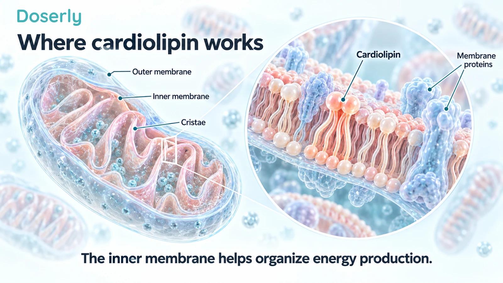
A mitochondrion has an outer membrane and a folded inner membrane. The folds, called cristae, provide an organized surface for proteins involved in energy production. These proteins do not operate in isolation: their surrounding lipids and arrangement influence how they work together.
Cardiolipin is one of those lipids. It contributes to the membrane environment and its interactions with proteins involved in respiration. In this context, respiration means the mitochondrial processes that use energy from fuels to support ATP production, rather than simply the movement of air through the lungs.
ATP transfers energy for cellular work. Producing it efficiently depends on a coordinated system, including electron transport and the membrane conditions needed to couple that transport to ATP synthesis. A problem in that system may involve the proteins, the lipids, their organization or other parts of the cell.
Early SS-31 experiments investigated cardiolipin because of this connection between membrane structure and function. They did not begin from the assumption that every symptom of fatigue is caused by a cardiolipin defect. Birk and colleagues, 2013
What the early binding experiments showed
A 2013 study used a fluorescent analogue to investigate SS-31's interaction with cardiolipin. The work connected that interaction with inhibition of a damaging peroxidase activity involving cytochrome c. In rats subjected to kidney ischemia, pretreatment was associated with preservation of mitochondrial membrane features and improved recovery of ATP-related processes. Original cardiolipin study
Ischemia means inadequate blood supply to tissue. The experimental problem was therefore a defined injury setting, not ordinary tiredness or healthy aging. The rat findings helped connect the membrane mechanism with tissue biology, but they did not establish clinical kidney protection in people.
A fluorescent analogue is also a research tool. It helps investigators observe an interaction; it isn't the same thing as measuring every consequence of the unmodified drug in a patient. The experiment's strength lies in identifying a plausible target and mechanism under controlled conditions.
This is a more useful explanation than saying SS-31 simply “finds damaged mitochondria.” That phrase leaves out what is being targeted, which experimental system was used and what changed.
Cytochrome c has more than one relevant behavior
Cytochrome c is a protein involved in electron transport. Its interaction with cardiolipin can also contribute to peroxidase activity, a chemical behavior associated with oxidation of lipids. The question was whether SS-31 could influence that interaction while preserving electron-transfer function.
A 2014 study examined model membranes, structural interactions and isolated mitochondrial preparations. The investigators reported that SS-31 interacted with cardiolipin-containing membranes and modulated the cytochrome c–cardiolipin relationship. Under the tested conditions, it preserved electron-carrier activity and improved aspects of respiration and ATP synthesis. Birk and colleagues, 2014
This helps explain why the mechanism is more specific than a generic “antioxidant” description. The experiments concern the membrane environment and a protein's behavior within it. They don't show that indiscriminately removing every reactive molecule would be beneficial, or that one mechanism explains every effect seen in different tissues.
The practical distinction is between altering a process and proving a patient outcome. A laboratory preparation can show improved electron transfer while leaving open whether an administered medicine will improve walking, fatigue or disease complications in people.

Does SS-31 “fix” cardiolipin?
The word “fix” can hide several different claims. It might mean changing how cardiolipin interacts with proteins, changing the amount or composition of cardiolipin, preventing a particular injury, or correcting the underlying genetic cause of a disease. These require different measurements.
Barth syndrome illustrates the distinction. It involves loss-of-function changes in TAFAZZIN, a gene involved in cardiolipin remodeling. Abnormal lipid composition is part of the disease biology. A peptide interacting with the membrane is not a gene replacement treatment.
In a 2022 study of tafazzin-knockdown mice, SS-31 improved cardiac mitochondrial respiratory capacity and organization of respiratory supercomplexes, groups of interacting proteins. The measured monolysocardiolipin-to-cardiolipin ratio did not change. The authors interpreted the findings as an effect on respiratory-chain function rather than direct correction of that lipid ratio. Russo and colleagues, 2022
That experiment is especially useful for the language question. Mitochondrial function improved in the model without the particular lipid abnormality being normalized. It would be inaccurate to describe the result simply as “cardiolipin repaired.”
What does the lipid ratio tell researchers?
Monolysocardiolipin, abbreviated MLCL, is related to cardiolipin remodeling. In Barth syndrome research, the MLCL/CL ratio is one way to characterize the lipid disturbance. It is a biochemical measurement, not a direct assessment of how far a patient can walk or how much force a muscle produces.
The long-term TAZPOWER extension reported changes in this ratio and associations with clinical outcomes in people with Barth syndrome. Ten participants entered the extension and eight reached week 168. The study had no concurrent placebo group during that period. TAZPOWER extension, 2024
The human extension and mouse experiment therefore should not be flattened into one universal statement about lipid correction. They involved different species, tissue measurements and treatment settings. A biomarker association in an uncontrolled extension also does not establish that changing that biomarker caused a functional improvement.
The useful question is which aspect of membrane biology changes, where it changes and whether that change explains the patient's outcome. The ratio is one part of that investigation, not a complete measure of mitochondrial health.
A ratio also combines two quantities. It can change because the numerator changes, because the denominator changes, or because both do. Reporting a lower MLCL/CL ratio therefore does not, by itself, tell the reader exactly which lipid pool changed or whether every relevant tissue responded similarly.
That is why the experimental details matter: which tissue was sampled, how lipids were measured and what functional assessment accompanied the result. A blood-based measurement and a measurement from isolated cardiac mitochondria are not automatically equivalent. The two studies are useful together because they ask related questions at different levels, not because one can be used as a direct replication of the other.
What is established in the prescription label?
The Forzinity prescribing information describes elamipretide as a mitochondrial cardiolipin binder that localizes to the inner mitochondrial membrane. The approved indication is improvement in muscle strength in adults and children with Barth syndrome weighing at least 30 kg, under accelerated approval. Forzinity prescribing information
That label is more specific than a general promise to repair mitochondria. Its clinical indication names the disease, the population and the intended outcome. It doesn't establish a benefit for everyone who experiences fatigue or believes their mitochondria need support.
The same label describes injection-site reactions and potentially serious hypersensitivity. A targeted mechanism does not eliminate adverse effects. Understanding where a medicine acts helps explain its development; it does not make the medicine automatically suitable or safe for every use.
Can the mechanism change energy production in human muscle?
A small randomized trial in 39 adults aged 60–85 selected for low mitochondrial function examined this question using a hand muscle. After one two-hour infusion, ATP-production capacity rose more in the elamipretide group on the percentage-change analysis. The absolute-change comparison narrowly missed conventional statistical significance, and there was no between-group difference at day seven. The prespecified fatigue-resistance analysis was not significant. Roshanravan and colleagues, 2021
This is direct human evidence about a biological measurement under selected conditions. It supports further investigation of mitochondrial effects, while showing why the mechanism should not be described as guaranteed sustained improvement.
It also separates capacity from experience. ATP-production capacity in one muscle and the feeling of having more energy are different outcomes. A physiological change may be useful, but researchers need to demonstrate the connection to daily function rather than assume it.
Why a plausible membrane target can coexist with a negative trial
MMPOWER-3 randomized 218 people with genetically confirmed primary mitochondrial myopathy to elamipretide or placebo for 24 weeks. Neither main outcome—six-minute walking distance or disease-specific fatigue—showed a significant benefit. MMPOWER-3, 2023
This does not invalidate every laboratory observation about cardiolipin. It shows that the expected clinical benefit did not emerge on those endpoints in that population and trial. A mechanism can be real while being insufficient to change the particular problem a person faces.
Several questions can explain why translation is difficult. Is the targeted process a major limitation in this disease? Does the relevant tissue receive enough exposure? Are other problems limiting function? Is the outcome sensitive to the kind of change produced? These are research questions, not reasons to reinterpret a negative result as a success.
The clinical finding should guide claims about benefit, while the mechanism helps guide the next hypothesis. Both are necessary for a complete account.
How to read a “mitochondrial repair” claim
Ask what was actually measured. Electron transport in isolated mitochondria, a lipid ratio, membrane appearance, ATP capacity and walking distance are all different observations. A claim should name the one the study supports.
Then identify the system: model membranes, cultured cells, rodents or people with a defined diagnosis. Human cells in a dish are not a human treatment trial. A positive animal injury experiment does not establish a preventive benefit in healthy adults.
Finally, distinguish modulation from replacement or cure. Elamipretide's interaction with cardiolipin does not mean it replaces mitochondria, edits TAFAZZIN or permanently normalizes every aspect of cellular energy production. Those are stronger claims requiring evidence that the cited studies do not provide.
What the membrane story is useful for
The cardiolipin research explains why elamipretide is more than an undirected idea about “boosting energy.” It identifies a location and a set of interactions that can be investigated experimentally. It also connects basic biology with a specific clinical-development program.
Its limits are equally informative. The mouse lipid-ratio finding, the mixed acute human ATP result and the negative myopathy primary endpoints show that different levels of evidence need to remain separate. They tell us where a simple repair narrative becomes too broad.
If you're discussing SS-31 because of symptoms or a diagnosed condition, ask how the proposed membrane effect relates to the outcome you need. The SS-31 guide brings together those clinical results and safety considerations, so the mechanism can inform the conversation without replacing it.
Sources
- Birk and colleagues, 2013: cardiolipin interaction and rat ischemia experiments.
- Birk and colleagues, 2014: model membranes, cytochrome c and mitochondrial preparations.
- Russo and colleagues, 2022: tafazzin-knockdown mouse mitochondrial function without lipid-ratio correction.
- TAZPOWER extension, 2024: human clinical and biomarker observations.
- Forzinity prescribing information: approved mechanism, indication and safety.
- Roshanravan and colleagues, 2021: selected older-adult ATP study.
- MMPOWER-3, 2023: randomized functional outcomes in primary mitochondrial myopathy.