
Peptides
October 16, 2026
Mazdutide and liver fat: does a better scan mean less scarring?
On this page
Mazdutide has produced substantial reductions in liver fat in human research. For someone living with fatty liver disease, that is an encouraging finding: excess fat is part of the disease process, and a treatment that changes it deserves careful study. But a lower liver-fat measurement doesn't by itself show that existing scar tissue has improved.
The distinction matters because liver disease has several dimensions. Fat accumulation, inflammation, liver-cell injury and fibrosis can be related without moving together at the same speed. A headline about an “80.2% reduction” can easily sound like the liver became 80.2% healthier. That isn't what the measurement says.
We'll examine where that mazdutide number came from, what an MRI can tell us, and what evidence would answer the separate question about scarring. Our mazdutide guide covers its broader metabolic and weight-management research.
Fat, inflammation and fibrosis are different findings
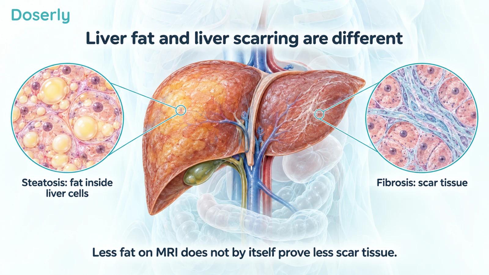
Steatosis means excess fat in liver cells. Metabolic dysfunction-associated steatotic liver disease, or MASLD, describes fatty liver associated with metabolic risk factors. Some people also develop inflammatory injury, called metabolic dysfunction-associated steatohepatitis, or MASH. Fibrosis is scar tissue that can accumulate in response to ongoing injury. Advanced scarring can progress to cirrhosis. NIH explanation of MASLD.
These terms describe different features of the liver, not interchangeable names for one scan result. A person may have considerable fat with little fibrosis, or fibrosis that remains clinically important after the amount of fat changes. Knowing that someone has “fatty liver” doesn't establish their fibrosis stage.
This is why clinicians consider medical history, blood tests and imaging together. Sometimes a biopsy is needed to examine tissue directly, but it isn't routine for every person with fatty liver. The appropriate assessment depends on the clinical question and the likelihood of more advanced disease. NIH MASLD information.
What did GLORY-1 actually study?
The main GLORY-1 trial was a weight-management study in 610 Chinese adults. Participants received weekly mazdutide 4 mg, mazdutide 6 mg or placebo for 48 weeks. Eligibility was based on body size and weight-related conditions, rather than requiring biopsy-confirmed MASH with a particular fibrosis stage. Its primary endpoints concerned body weight. GLORY-1 primary report.
That doesn't make liver measurements unimportant. It does mean that a liver-fat subgroup analysis answers a different question from a trial designed specifically to test whether a medicine improves MASH or fibrosis.
In a June 2024 sponsor announcement, Innovent described an exploratory analysis of participants with MRI measurements. Ninety-two had a baseline scan; 69 had liver fat of at least 5% at baseline and an assessment at week 48. They included 25 assigned 4 mg, 22 assigned 6 mg and 22 assigned placebo. Original Innovent announcement.
The sponsor reported mean relative changes in liver fat of −63.3%, −73.2% and +8.2%, respectively. The 80.2% reduction in the headline applied specifically to the 6-mg subgroup starting with liver fat of at least 10%. It was not the average result for all 610 randomized participants or a measurement of scar-tissue reduction. The source was a company report of exploratory findings, rather than a standalone peer-reviewed fibrosis trial.
That leaves a useful and appropriately limited conclusion: mazdutide reduced MRI-measured liver fat in the assessed subgroup. The fibrosis question requires a different outcome.
What does “relative reduction in liver fat” mean?
MRI-PDFF stands for magnetic resonance imaging proton density fat fraction. It estimates the proportion of the relevant MRI signal attributable to fat relative to fat and water. It is useful for quantifying liver fat and following changes without repeatedly removing tissue. It doesn't measure the percentage of the organ that has healed. AASLD practice guidance.
A numerical example makes the distinction clearer. Imagine a liver-fat measurement falling from 20% to 4%. That is a decrease of 16 percentage points. Relative to the starting value, it is an 80% reduction, because 16 divided by 20 equals 0.80. These are hypothetical numbers chosen to explain the arithmetic, not individual GLORY-1 participant data.
Now imagine a change from 10% to 2%. It is also an 80% relative reduction, although the absolute decrease is eight percentage points. The starting value matters when interpreting relative percentages.
Neither example contains information about fibrosis. A separate scar-tissue assessment could improve, remain unchanged or show a more complicated pattern. It would have to be measured; the fat calculation cannot supply it.
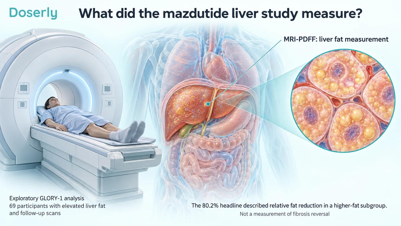
Image sources: Source
Why the subgroup deserves attention
An imaging substudy can reveal a meaningful biological effect without including every trial participant. MRI takes time and resources, and not every center or participant may be included in a scanning program. The critical issue is to keep the analysis population visible.
Here, “69 participants with elevated liver fat and follow-up scans” is more informative than “a phase 3 study of 610 people.” The larger number describes the parent trial. The smaller number describes the exploratory analysis producing the liver-fat results. They shouldn't be substituted for one another.
Requiring a follow-up measurement also raises a practical question: how similar were the people with usable scans to those without them? If missing scans are related to treatment tolerance or response, a complete-case subgroup can differ from the full randomized population. That is a potential limitation, not a claim that the observed result is wrong.
For the same reason, a higher-fat subgroup may have more room for measurable improvement. Its result is useful for generating and testing a focused liver-disease hypothesis. It should not be generalized to everyone with obesity, everyone taking the drug or everyone with established cirrhosis.
Does newer mazdutide research change the conclusion?
A later Innovent announcement described GLORY-2, a 60-week study of 462 Chinese adults with obesity, including some participants with diabetes. In the MRI subset without diabetes and with baseline liver fat of at least 10%, the sponsor reported a 71.9% relative decrease with mazdutide 9 mg versus a 5.1% increase with placebo. GLORY-2 sponsor report.
This adds another liver-fat signal, under a different regimen and set of study conditions. It still doesn't convert MRI-PDFF into a fibrosis measurement. The report describes a selected imaging subgroup, and its company-announcement status should remain attached to the numbers.
You also cannot use the 80.2% and 71.9% figures to decide that one dose is better for the liver. They come from different studies and subgroup definitions. A direct dose comparison would need comparable participants, measurements, follow-up and analysis.
The repeated direction of liver-fat change makes continued liver research reasonable. The next question is whether that change translates into improvements in inflammatory injury, fibrosis and outcomes that matter over years.
What about ALT and other blood tests?
ALT, alanine aminotransferase, is an enzyme measured in blood. Its level can rise with liver-cell injury. A falling ALT can be encouraging, but it is not a direct measurement of collagen or scar tissue. The GLORY-1 publication reported improvements in prespecified cardiometabolic measures; those results should retain their individual names rather than being compressed into “liver recovery.” GLORY-1.
A normal enzyme result also doesn't establish that clinically important liver disease is absent. AASLD guidance emphasizes that aminotransferases should not be used alone to exclude advanced disease. Fibrosis assessment requires a broader evaluation. AASLD guidance.
This distinction can help when your own results appear inconsistent. A clinician may be pleased with lower liver fat or enzymes while still recommending fibrosis assessment or ongoing follow-up. That isn't necessarily a contradiction. The tests are measuring different aspects of the problem.
What would demonstrate fibrosis improvement?
A fibrosis study needs to identify the participants' starting disease and use an outcome that actually assesses scarring. Liver biopsy permits examination of tissue architecture and fibrosis stage, although it samples only part of the liver and has limitations. Noninvasive fibrosis assessments can also be clinically useful, but their interpretation depends on the test and the context.
FDA's original review of resmetirom, a different medicine, illustrates the separation between liver trial endpoints. The approval evidence included biopsy-based assessments of steatohepatitis resolution without worsening fibrosis, and fibrosis improvement without worsening steatohepatitis. These are two distinct outcomes, even though they concern the same disease. FDA trial snapshot.
That example does not supply missing mazdutide evidence. It shows what it means to measure an outcome specifically. Even biopsy endpoints can be surrogates: measurements expected to predict future clinical benefit rather than direct observations of fewer liver failures or deaths. Longer follow-up is needed to assess those outcomes.
A persuasive mazdutide fibrosis claim would therefore identify the starting fibrosis stage, the comparator, the assessment method, the response definition, the number assessed and the uncertainty around the result. An MRI fat reduction alone cannot answer those questions.
How to discuss an improving scan with your clinician
If your liver-fat measurement falls, ask what changed and what remains to be assessed. It may be a meaningful response while still leaving a separate question about fibrosis. Keep the test name, date and units with the result; “my liver score improved” can hide whether the measurement was fat fraction, stiffness or a blood-based estimate.
Ask how your fibrosis risk was assessed and whether that assessment changes the follow-up plan. Also discuss alcohol intake, metabolic conditions and other medicines, because a liver finding doesn't exist separately from the rest of your health. The aim is to understand your actual disease and care plan, not to order every possible test because a research article mentioned it.
For mazdutide, the current findings discussed here support a liver-fat effect in studied populations. They don't prove fibrosis reversal. That distinction allows the promising result to remain promising without giving it a different clinical meaning. The mazdutide evidence section provides context on the compound's other human outcomes.
Explore the research
- GLORY-1 primary publication, 2025: parent weight-management trial.
- Innovent exploratory liver-fat report, June 2024: subgroup definitions and the 80.2% headline.
- GLORY-2 sponsor announcement: later MRI subset findings, not a peer-reviewed fibrosis report.
- NIH MASLD explanation: disease terminology and assessment.
- AASLD practice guidance, 2023: MRI-PDFF, enzymes and fibrosis assessment.
- FDA Rezdiffra trial snapshot: an example of distinct biopsy endpoints for another medicine.