
Supplements
October 16, 2026
Vitamin K2 and calcium: what the routing diagram leaves out
On this page
The familiar vitamin K2 diagram is persuasive: calcium moves into bone and stays out of arteries. It compresses real biology into a simple visual promise. The problem is that a nutrient's role in activating proteins does not establish that a supplement will strengthen bones, reverse arterial deposits or prevent a heart attack.
Human research is more interesting than either an enthusiastic sales pitch or a blanket dismissal. Bone trials have produced mixed results. An older aortic-valve trial was negative, while two coronary-artery trials published in 2026 reported slower progression of calcification on imaging. The meaning depends on the tissue, product, population and outcome.
The useful question is therefore specific: what did a particular K2 intervention change, and did that change improve health in a way the study actually measured?
The biology is real, but it is not a plumbing system
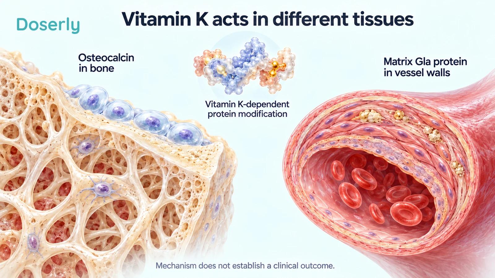
Vitamin K helps enzymes modify certain proteins through a process called carboxylation. Among the proteins involved are osteocalcin in bone and matrix Gla protein, often abbreviated MGP, in tissues including blood vessels. This provides a biological rationale for studying vitamin K in bone and vascular health. NIH vitamin K fact sheet
That mechanism does not mean K2 physically collects calcium from an artery and delivers it to a bone. Bone mineralization and vascular calcification are distinct biological processes. A diagram drawing one arrow between them can accidentally imply a transfer that a clinical trial has not demonstrated.
The distinction matters even when a supplement changes a relevant protein measurement. A lower amount of an undercarboxylated protein can show that vitamin K affected a biochemical pathway. It does not, by itself, show that a person avoided a fracture or cardiovascular event.
Our vitamin K2 guide provides more background on forms and uses. Here, the focus is how far the clinical evidence supports the common calcium-routing claim.
K2 is a family, not one interchangeable treatment
Vitamin K1 is phylloquinone. Vitamin K2 refers to menaquinones, including MK-4 and MK-7. Supplements and trials use different forms and amounts, and those distinctions should remain visible when discussing results. NIH vitamin K forms
For example, an MK-4 trial using milligram quantities does not directly test an MK-7 product labeled in micrograms. One milligram equals 1,000 micrograms, but unit conversion alone does not make different compounds clinically equivalent.
Combination products create another complication. If a trial gives K2 together with vitamin D, its result applies to that combination compared with the control. It cannot isolate how much of the effect came from K2. If both groups already receive calcium and vitamin D, the question is what adding vitamin K contributes on top of that background regimen.
These are not technicalities to ignore after reading the headline. They define the intervention. A useful comparison starts by matching the exact form, amount, co-ingredients and duration before discussing whether a commercial product resembles the study.
A classic bone trial changed the marker without changing density
A 2009 double-blind trial enrolled 381 healthy postmenopausal North American women without osteoporosis. Participants received vitamin K1 at 1 mg daily, MK-4 at 45 mg daily or placebo for 12 months. Everyone also received calcium and vitamin D3. Binkley bone trial
The vitamin K groups had lower undercarboxylated osteocalcin, a biochemical response consistent with the mechanism. But the investigators did not find improvements in lumbar-spine or proximal-femur bone mineral density, selected bone-turnover markers or femoral geometry.
This is a particularly useful study because it tests the leap in the routing diagram. The pathway responded, yet the measured structural outcomes did not follow over that year. That does not prove all vitamin K interventions fail in every population. It shows why “activates osteocalcin” cannot be used as shorthand for “builds stronger bones.”
It also was not an MK-7 trial. Using it to dismiss every MK-7 product would blur the same form-specific distinction that matters when interpreting favorable studies.
MK-7 bone studies have not all agreed
A 2013 study randomized 244 healthy postmenopausal women to 180 micrograms of MK-7 daily or placebo for three years. It reported less decline in bone mineral density at the lumbar spine and femoral neck, but not at the total hip, along with changes in some calculated bone-strength measures. Three-year MK-7 study
Those are encouraging structural findings in that population. They should be described at the sites that improved, without expanding the result to every skeletal measurement or claiming that the trial established fewer hip fractures.
A separate three-year trial, published in 2021, enrolled 142 postmenopausal women with osteopenia. It tested 375 micrograms of MK-7 daily as an addition to calcium and vitamin D, which both groups received. Despite a substantial change in osteocalcin carboxylation, the study did not find a between-group advantage in bone mineral density or the assessed bone microarchitecture. Osteopenia add-on trial
The studies differed in population, regimen and background supplementation. Their disagreement is not resolved by picking the larger dose or treating every vitamin K marker as a bone-strength result. For someone with low bone density, the evidence needs to fit inside an osteoporosis assessment rather than replace it.
An aortic valve is not a coronary artery
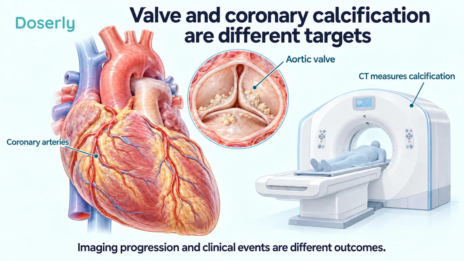
Calcification in a heart valve and calcification in the arteries supplying the heart are related clinical topics, but they are different anatomical targets. A supplement can be tested against one without establishing its effect on the other.
The 2022 AVADEC trial studied 365 men with aortic-valve calcification. Participants received 720 micrograms of MK-7 plus 25 micrograms of vitamin D daily, or placebo, for two years. The intervention did not significantly slow the primary outcome of aortic-valve calcification progression. AVADEC trial
The result is relevant to claims about preventing or treating valve calcification with that regimen. It should not be relabeled as proof that every vascular use of K2 is ineffective. Equally, a favorable result in an artery cannot be used to claim that a calcified valve has been repaired.
This is one reason an illustration of a generic “clean artery” is a poor substitute for the actual trial question. It hides both the anatomical site and the difference between slowing a process and reversing an existing abnormality.
What the newer coronary trials add
The VitaK-CAC trial, published in 2026, randomized 180 patients with symptomatic coronary artery disease and baseline calcium scores between 50 and 400 Agatston units. Participants were assigned 360 micrograms of MK-7 daily or placebo for two years; the report describes 85 and 82 patients receiving the respective interventions. VitaK-CAC trial
Coronary calcium scores increased in both groups, but the increase was smaller with MK-7. This supports an effect on the imaging outcome in the studied population. It does not show that existing calcium disappeared. The investigators explicitly left the clinical implications for plaque stability unresolved.
A second 2026 trial, DANCODE, addressed more severe coronary calcification. It randomized 400 participants with calcium scores of at least 400; 398 were included in the report. The intervention combined 720 micrograms of K2 with 25 micrograms of D3 daily for 24 months. DANCODE trial
The mean calcium-score increase was 196 Agatston units with supplementation and 248 with placebo, a between-group difference of 52 units favoring the intervention. A plaque-imaging subset did not show an increase in noncalcified plaque volume attributable to treatment. These are meaningful additions to the evidence, but the tested product was a combination and the primary outcome was imaging progression.
Taken together, these newer studies make a simple “K2 has never affected arterial calcification” statement outdated. They still do not establish that a consumer taking a D3/K2 product will have fewer heart attacks, live longer or move calcium from arteries into bone.
Slower progression, reversal and fewer events are separate claims
Imagine two groups whose imaging scores both rise, but one group's score rises less. Calling that result “slower progression” preserves what happened. Calling it “decalcification” suggests that deposits shrank, which is a different result.
The same discipline applies to the next step. A marker may predict risk in an observational study without being a proven treatment target. Demonstrating that an intervention changes the marker does not automatically demonstrate that changing it improves clinical outcomes. The VitaK-CAC investigators' caution about plaque stability makes this particularly relevant here. VitaK-CAC interpretation
This does not make imaging research unimportant. Imaging can show whether an intervention affects a process and help design the next trial. It simply means the result should be reported at the level it reached.
For a reader comparing product claims, the practical test is to identify the noun after “improved”: a vitamin K marker, a bone-density site, arterial stiffness, a calcification score, a fracture rate or a cardiovascular-event rate. Those nouns name different outcomes. A confident statement about one should not silently substitute for evidence about another.
K2 does not turn a high-dose supplement stack into a proven safe plan
Pairing vitamin D, calcium and K2 can sound as though each ingredient protects against the risks of the others. The trials above do not establish that K2 neutralizes excessive calcium or vitamin D exposure. They tested specified regimens in specified populations; they did not provide a universal insurance policy for larger doses.
A combination bottle also makes it easier to overlook duplication. A separate vitamin D product, a multivitamin and a bone supplement may all contribute ingredients. Before interpreting the K2 amount, identify the total calcium and vitamin D supplied by the routine. The calcium guide and vitamin D3 guide can help clarify those separate ingredients.
The reason for taking each nutrient should remain understandable without the marketing diagram. If the only explanation is that one ingredient is needed to make another unnecessary high dose safe, the plan deserves a fresh review.
The anticoagulant interaction needs an explicit check
Vitamin K can meaningfully interact with warfarin and related vitamin K antagonists. Consistent intake matters, and starting or stopping a K2 supplement can change the situation the anticoagulation regimen was designed around. NIH advises maintaining a consistent vitamin K intake when using these medicines. NIH vitamin K interactions
This is not an instruction to remove all vitamin K foods, and it is not a claim that every anticoagulant works through vitamin K. The exact medicine matters. Bring the product label and intended change to the clinician or pharmacist responsible for anticoagulation rather than making the change first and mentioning it later.
The issue also illustrates why “no upper limit established” should not be interpreted as “no clinically important constraints.” An interaction can matter even when a nutrient is ordinarily well tolerated.
Make the record match the question
A useful supplement record includes MK-4 or MK-7, the amount and units, other ingredients, start date and the intended reason for use. If a clinician is following bone density or cardiovascular disease, record the existing treatment plan and the relevant review date as well.
Doserly can help keep that history together. It cannot determine whether an imaging change represents clinical benefit or whether a supplement should replace an established treatment.
The most accurate description of K2 is neither a calcium traffic controller nor a biologically irrelevant vitamin. It participates in important pathways, and trials have tested some promising effects. The decision becomes clearer when the mechanism, measured outcome and remaining uncertainty stay separate.
Sources
- NIH Office of Dietary Supplements: Vitamin K
- Binkley and colleagues: Vitamin K, osteocalcin and bone outcomes, 2009
- Knapen and colleagues: Three-year MK-7 bone trial, 2013
- Rønn and colleagues: MK-7 in osteopenia, 2021
- Diederichsen and colleagues: AVADEC, 2022
- Vossen and colleagues: VitaK-CAC, 2026
- Hasific and colleagues: DANCODE, 2026