
Peptides
October 16, 2026
Visceral and subcutaneous fat: why the distinction matters
On this page
Two people can have a similar waist measurement and different distributions of abdominal fat. One may carry more fat beneath the skin, another more inside the abdominal cavity. Both tissues contribute to body composition, but their location, relationship to metabolic risk, and response to a treatment are not interchangeable.
That is why the phrase “belly fat” can become misleading in a medication discussion. Tesamorelin trials, for example, distinguished visceral adipose tissue from abdominal subcutaneous adipose tissue. A favorable result in the first compartment did not establish the same result in the second. Understanding the anatomy helps explain why a scan, a tape measure, and a bathroom scale can tell different stories. Pooled tesamorelin trials
The distinction should help people interpret their health information, not encourage them to diagnose an internal fat compartment by appearance. A visible abdomen is not a metabolic assessment, and an apparently lean body does not establish that every relevant health marker is favorable.
Start with the abdominal wall
Subcutaneous means beneath the skin. In the abdomen, this fat lies outside the muscular wall. Visceral fat occupies deeper spaces within the abdominal cavity, around organs such as the bowel. The muscle wall provides a useful anatomical boundary when looking at an imaging cross-section.
A skinfold mainly samples skin and some underlying subcutaneous tissue. It does not reach through the abdominal wall to measure visceral fat. Similarly, a person’s impression that their abdomen feels soft or firm cannot reliably quantify the internal compartment. Muscle tension, posture, digestive contents, and other factors can change what the abdomen looks or feels like.
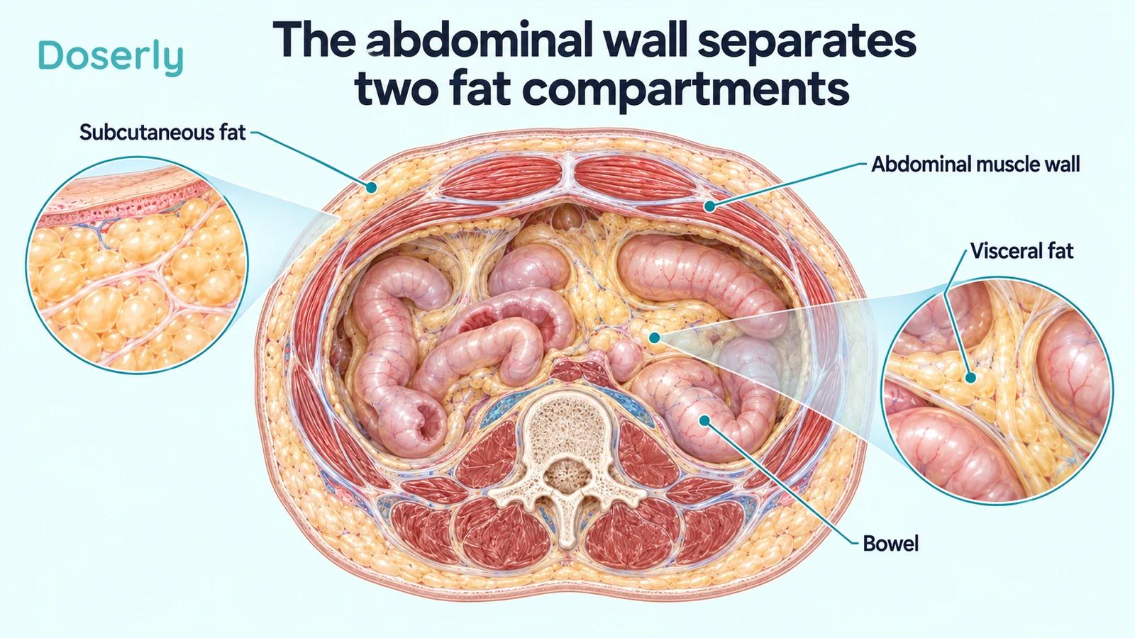
This is also why fat within the liver deserves a separate name. Liver fat refers to fat stored in liver tissue, while visceral adipose tissue is a fat depot outside that tissue. The two may be related, but a visceral-fat measurement does not directly quantify hepatic fat. In tesamorelin research, investigators used different imaging methods to assess those outcomes. Randomized liver-fat trial
Calling all three “internal fat” may be convenient in conversation, but it loses information when discussing results. The more useful question is which compartment a test or study actually assessed.
Why visceral fat attracts clinical attention
Community studies have found that visceral fat carries information about metabolic risk beyond overall body size. In a Framingham Heart Study analysis of 3,001 adults, both visceral and subcutaneous abdominal fat were associated with metabolic risk factors, but visceral fat had stronger relationships with most of them. Its associations remained informative after accounting for body mass index and waist circumference. Framingham compartment analysis
This does not make subcutaneous fat biologically inactive or uniformly harmless. It means that the amount and location of fat can contribute different information. A simple “bad fat versus good fat” label is too crude to describe the findings, particularly when a person’s overall health depends on many factors beyond a single tissue depot.
Longitudinal research adds another perspective. A Framingham analysis followed 1,730 participants for an average of 6.2 years and found that baseline fat measures were associated with subsequent changes in metabolic risk factors. Visceral-fat associations were generally more pronounced than those for subcutaneous fat. Because this was observational research, it did not test whether a particular drug-induced reduction would produce the same change in risk. Prospective metabolic-risk study
That distinction matters whenever a treatment advertisement jumps from “visceral fat is associated with disease” to “this treatment prevents disease.” The first statement helps explain why researchers study the tissue. The second requires treatment-outcome evidence. A medicine can reduce a risk-associated measurement without yet having demonstrated fewer heart attacks, less disability, or longer survival.
Similar body size can conceal different distributions
Body mass index uses weight and height. It is useful for many population and clinical purposes, but it does not identify where fat is stored or distinguish fat from lean tissue. Waist circumference adds information about abdominal size, yet it still measures around all the tissues inside that perimeter.
A Framingham study of 3,348 adults examined patterns of visceral and subcutaneous fat using CT. More than a quarter of participants had discordant classifications for high visceral versus high subcutaneous fat under the study’s definitions. The authors found that BMI and waist circumference could misclassify people in relation to visceral fat and metabolic risk. Those research cutoffs are not universal self-diagnosis thresholds. Patterns of abdominal fat distribution
An everyday implication is that comparing waist sizes between friends is not a reliable comparison of metabolic health. It also means that a stable waist does not prove every underlying compartment is stable. Changes can be small, distributed differently, or offset by changes in other tissues.
None of this makes home measurements pointless. Repeated weight or waist measurements can help describe a trend when the method is consistent. They simply answer a broader question than an imaging assessment that separates tissues. Their value increases when they are interpreted alongside the person’s clinical history, symptoms, and relevant laboratory measurements.
What a scan measures—and what its units mean
A CT or MRI analysis can distinguish anatomical compartments in ways a tape measure cannot. Some research, including the tesamorelin trial's CT assessment, reports an area from a cross-section, commonly in square centimetres. Other research estimates a volume across multiple slices, commonly in cubic centimetres. Those units describe different quantities and should not be substituted for one another.
Imagine a stack of thin slices through an object. One slice can show the area of a particular tissue at that level; adding information across many slices can estimate its volume. The relationship depends on the shape and distribution of the tissue. Multiplying a single area by an arbitrary number does not produce a validated whole-abdomen measurement.
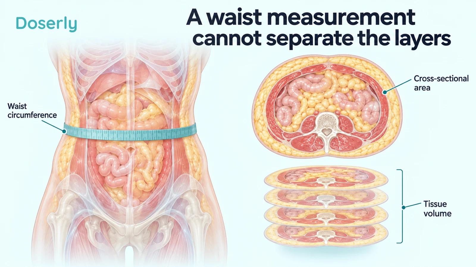
This helps when comparing study headlines. A reduction of a certain number of square centimetres is not a reduction of that many centimetres around the waist. A percentage reduction in visceral-fat area is not the same percentage reduction in total body fat or body weight. The denominator is part of the result.
It is also important to compare like with like over time. Different scan levels, analysis methods, or devices may not be directly interchangeable. A clinician interpreting repeated measurements needs to know how they were obtained, not just whether the later number is lower. Imaging may be appropriate for some clinical questions, but the existence of a precise research measure does not mean everyone should seek repeated scans.
Tesamorelin shows why compartment-specific wording matters
In the pooled phase 3 tesamorelin analysis, the reported 26-week visceral-fat treatment effect was approximately −15.4% relative to placebo. Abdominal subcutaneous fat did not show a statistically significant between-group effect. The participants were adults with HIV and excess abdominal fat receiving antiretroviral therapy. Pooled randomized trials
The accurate interpretation is a selective finding within that studied setting. It is not evidence that every layer of abdominal fat shrinks equally. It also does not mean subcutaneous fat can never change in an individual receiving treatment; the group comparison simply did not establish that effect in the study.
The US label describes tesamorelin as weight neutral and does not indicate it for weight-loss management. That is compatible with an internal compartment changing while total weight remains similar. It is also why a person seeking substantial overall weight loss needs a different discussion from someone being evaluated for HIV-associated lipodystrophy. Current EGRIFTA WR label
Doserly’s tesamorelin evidence guide describes the medication studies in more detail. The anatomical lesson applies more broadly: a treatment claim should name the tissue, the measurement, and the clinical population rather than rely on the visual familiarity of the word “belly.”
Losing fat is not always the same clinical goal
In HIV care, fat accumulation and fat loss can coexist in different regions. NIH guidance describes lipodystrophy as including both lipohypertrophy, or regional accumulation, and lipoatrophy, or loss of subcutaneous fat, particularly from the face, buttocks, and limbs. Prior treatment history can remain relevant even after the implicated medicines have changed. NIH guidance
For someone already troubled by loss of tissue in those regions, “more fat loss” is not a sufficiently specific treatment goal. The desired change may concern an internal abdominal compartment while preserving tissue elsewhere. This is another reason a generalized weight-loss headline can fail to describe the person’s actual concern.
It also changes the interpretation of body shape. A more prominent abdomen can be perceived differently when the limbs or face have lost fat. Clinical assessment needs the pattern across regions and the history over time, rather than treating one photograph or one measurement as the entire problem.
Fat inside muscle is another distinct question
Muscle imaging sometimes reports tissue density as well as area. This can provide information about fat within muscle, but it is not the same as measuring the visceral compartment. Nor does a change in muscle appearance on imaging automatically establish improved strength, mobility, or endurance.
An exploratory tesamorelin analysis assessed trunk-muscle CT measures after 26 weeks. Its treated group was restricted to participants who had already achieved a visceral-fat reduction of at least 8%. Muscle area and density improved on several measures, but the authors called for research into daily-life consequences and longer-term effects. Exploratory muscle analysis
Selecting responders makes that analysis different from asking what happens to everyone who starts treatment. It can help investigate accompanying tissue changes in those who respond, but it cannot establish an average functional benefit for an unselected population. For a person whose main goal is easier stair climbing or better strength, those functions should be assessed directly rather than inferred from a scan.
A smaller compartment is not a complete risk assessment
Visceral fat is one part of a larger clinical picture. Blood pressure, glucose regulation, lipids, smoking, family history, medications, and existing disease all influence decisions. A favorable imaging change does not establish that those other factors are controlled, just as an unfavorable single measurement does not determine a person’s entire future.
The tesamorelin label makes this particularly concrete: long-term cardiovascular safety has not been established. Its warnings about glucose intolerance and elevated IGF-1 remain relevant even when visceral fat falls. A treatment can have a desired tissue effect and still require monitoring for effects elsewhere. Prescribing information
For people with HIV, the broader care plan also needs to distinguish general weight gain from a regional fat-distribution problem. The current NIH guidance discusses these issues within ongoing HIV care rather than treating a single body-composition measurement as a reason to interrupt effective therapy. NIH guidance on weight gain in treated HIV
Reading a result without overreading it
When a report or clinic describes a change in abdominal fat, four details make the statement more useful: which compartment was measured, which method was used, what the units represent, and which outcome the result is supposed to inform. “Visceral-fat area on CT” is much clearer than “fat loss,” even if the second phrase sounds more appealing.
A follow-up discussion can then connect the measurement to the person’s goals. If the goal is a change in abdominal discomfort, appearance, metabolic health, or overall weight, ask how that goal will be assessed separately. A single test may contribute useful information without answering every question.
Keeping the compartments distinct is not technical fussiness. It prevents a result from being enlarged as it moves from a research paper to a headline or a treatment conversation. Subcutaneous fat, visceral fat, liver fat, and muscle fat describe different tissues. Understanding which one changed makes the evidence more useful—and makes room for a more accurate account of what remains unknown.
Sources
- Fox and colleagues: abdominal fat compartments and metabolic risk.
- Prospective Framingham study of fat depots and metabolic-risk changes.
- Patterns of abdominal fat distribution in Framingham.
- Pooled phase 3 tesamorelin trials.
- Randomized tesamorelin liver-fat trial.
- Exploratory tesamorelin muscle analysis.
- EGRIFTA WR prescribing information.
- NIH guidance on weight gain in treated HIV.