
Peptides
October 16, 2026
What immune-cell markers can reveal, and what they leave unanswered
On this page
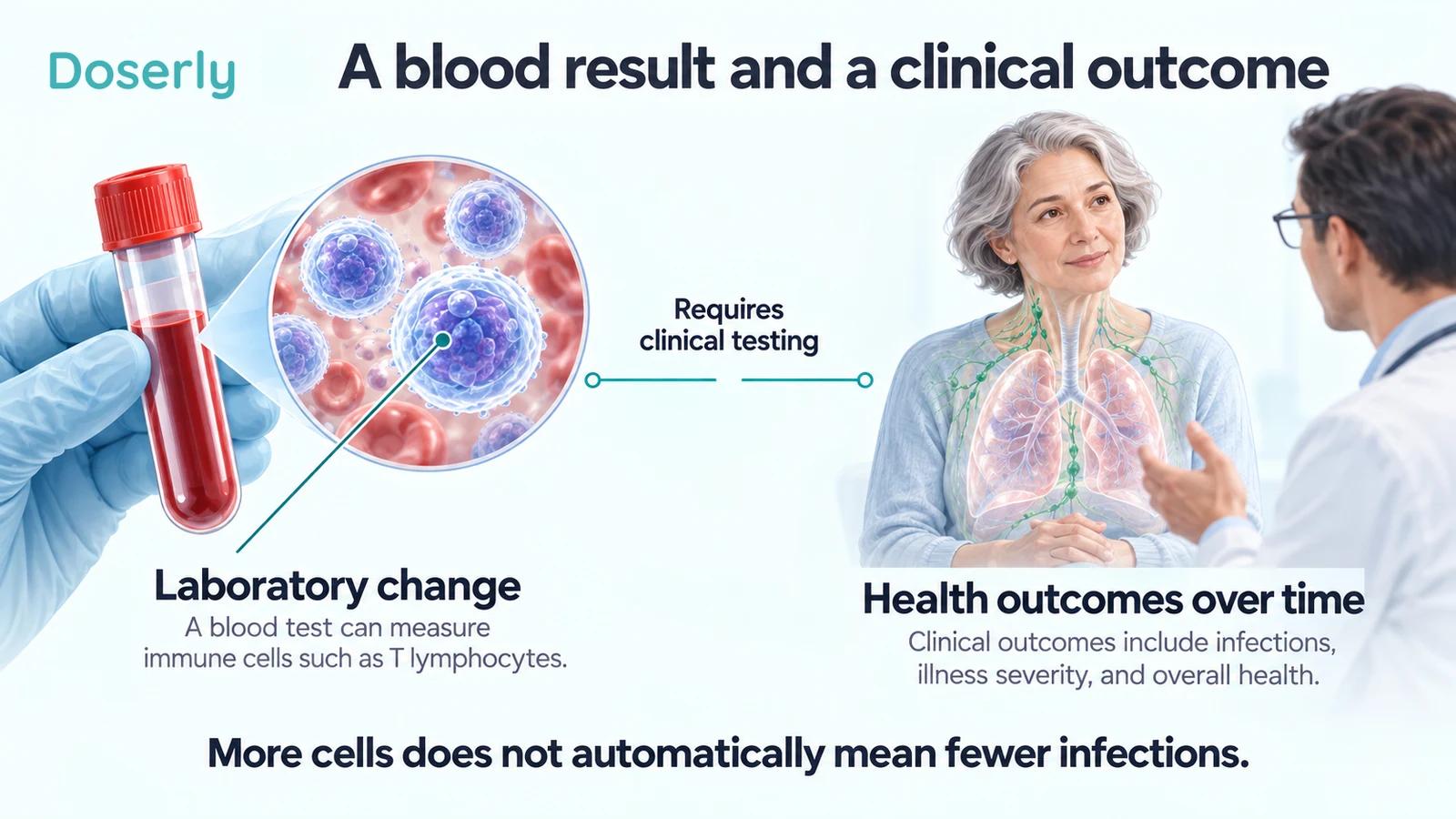
An immune-cell count rises after treatment. A protein associated with cell exhaustion becomes less common. A laboratory test suggests that more newly formed T cells are entering circulation. These can be useful findings, but they do not all mean the same thing—and none automatically tells us that a person will have fewer infections or live longer.
Thymosin alpha-1 research makes these distinctions especially relevant. Studies have measured cell numbers, cell subsets, surface proteins and markers related to thymic output. Some findings suggest biological activity. The harder question is whether changing those measurements improves an outcome patients can feel or experience.
Understanding the difference does not require dismissing laboratory research. It requires knowing what each measurement can support, how it was compared and whether the study followed people long enough to answer the clinical question.
A cell count is one view of a complicated system
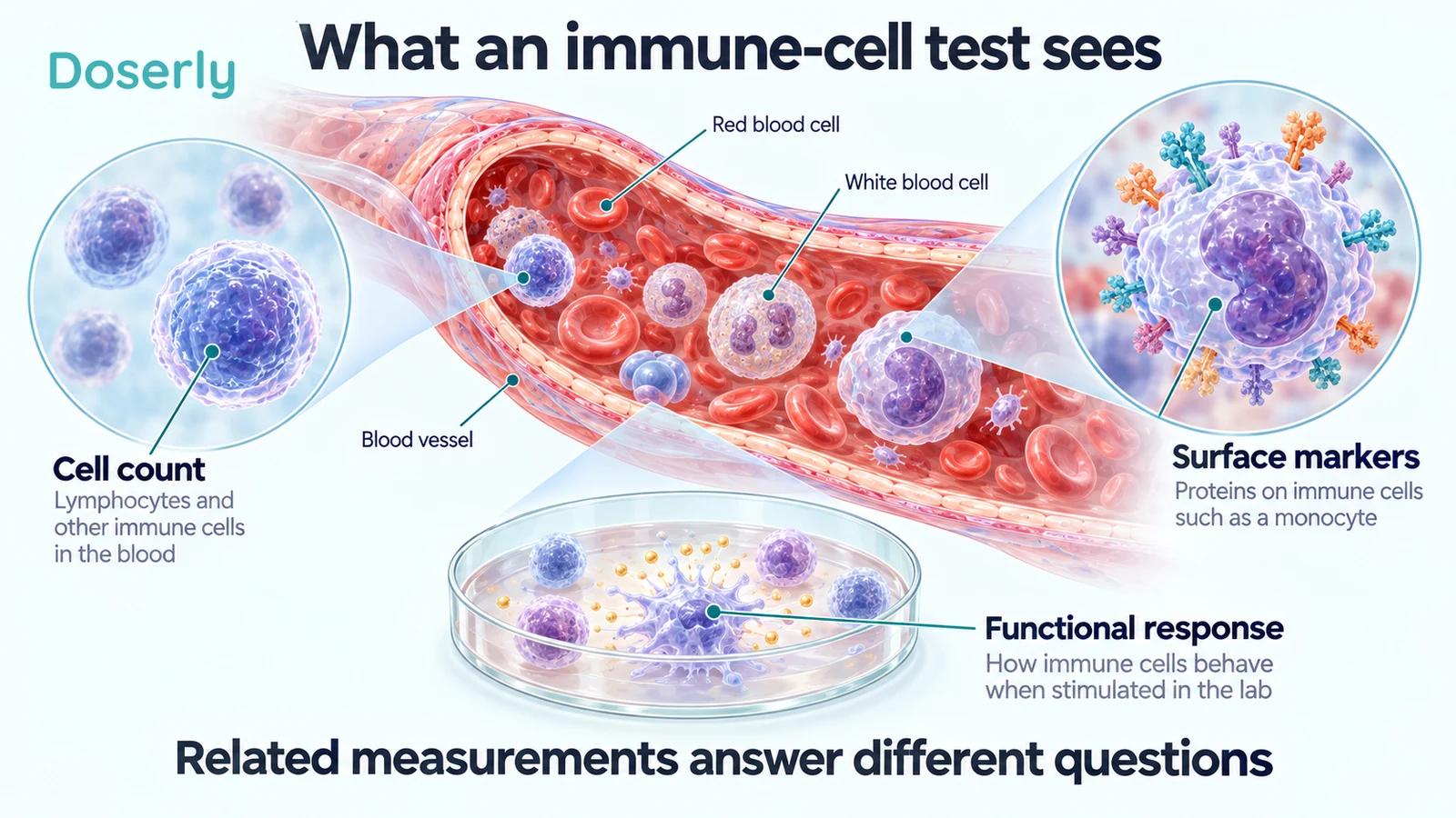
A count tells us how many cells meeting a definition were found in a specified amount of blood. A CD4 T-cell count, for example, counts a particular population of lymphocytes. A percentage answers a different question: what share of the measured population belongs to that group?
That distinction can matter even before considering treatment. Imagine a purely illustrative sample containing 100 cells, of which 20 belong to a subgroup. The subgroup accounts for 20%. If the total falls to 50 while the subgroup remains at 20, its percentage rises to 40% without any increase in its absolute number. Real laboratory calculations are more complex, but the denominator problem is the same.
Cell populations also contain different subsets. A study might distinguish naïve cells from memory cells, or measure a protein on the surface of cells rather than counting the cells themselves. Reading all these results as “immunity increased” erases the information that makes the findings interpretable.
The 2024 thymosin alpha-1 study in people with incomplete immune recovery despite HIV treatment is a useful example. It measured several such outcomes, which did not all move in the same way.
What the small HIV study actually found
The 2024 study treated 20 people while they continued antiretroviral therapy. It had one treatment group, without a randomized untreated comparison group. At 24 weeks, the investigators reported changes in selected T-cell proportions and lower expression of PD-1, a protein studied in relation to immune exhaustion.
However, absolute CD4 counts and a marker of thymic output did not show significant differences from baseline at the final 24-week endpoint. TIM-3, another measured surface protein, also did not change significantly at that endpoint. Earlier time-point findings and final endpoint findings therefore need to remain distinct.
This is more informative than saying simply that the treatment “restored immunity.” Different measurements produced different results, and the study design leaves uncertainty about what would have happened without the added treatment. It supports further investigation of specific biological effects. It does not establish that the participants became less likely to develop opportunistic infections or other illnesses.
There is also a difference between observing lower expression of one exhaustion-associated marker and demonstrating that the entire immune response has recovered. A marker contributes evidence about a cell state; it is not a complete functional examination of the immune system.
Thymic-output markers are indirect evidence
An earlier 2003 randomized pilot in 20 people with HIV examined thymosin alpha-1 alongside existing antiretroviral therapy over 12 weeks. It found a change in T-cell receptor excision circles, commonly abbreviated TRECs, without a significant between-group improvement in CD4 counts.
TRECs are small DNA circles generated during the rearrangement of T-cell receptor genes. Researchers use them as an indirect window into the contribution of newly generated T cells. They do not count prevented infections, and their interpretation is not identical to measuring the number or performance of all circulating T cells.
The pilot is therefore valuable for a specific reason: it suggests that a biological pathway may be affected even when a familiar cell-count endpoint does not show the expected change. That can help design subsequent studies. It cannot answer every downstream question at once.
If a summary mentions only the favorable TREC finding, a reader loses the accompanying CD4 result and the small study size. Good interpretation keeps those facts together. A signal that helps explain mechanism can be real and still fall short of a treatment recommendation.
Why association does not guarantee a treatment target
Some immune measurements are associated with prognosis. People with a particular abnormal result may, on average, have a higher risk of illness. That makes the measurement potentially useful for understanding risk, but it does not prove that deliberately moving the number will reduce that risk.
The distinction is causal. A marker may participate in disease, reflect disease severity or change alongside other processes. An intervention may alter the marker while leaving the most important disease mechanisms unaffected. It may also have other effects that offset a favorable laboratory change.
A striking human example comes from a different treatment, interleukin-2. The SILCAAT and ESPRIT randomized trials enrolled 1,695 and 4,111 people with HIV, respectively. Adding interleukin-2 produced sustained increases in CD4 counts, yet did not reduce the trials’ primary clinical outcome of opportunistic disease or death.
Those results do not prove that every immune-directed therapy fails. They demonstrate why a treatment-induced increase in a prognostic marker needs its own clinical validation. A measurement can be important in routine care without being a reliable substitute for outcomes when evaluating a new intervention.
Sepsis studies ask a different immune question
In sepsis, researchers have studied monocyte HLA-DR expression as one indicator of altered immune function. This is a surface-protein measurement on a different cell population, not another name for a CD4 count.
The 2013 ETASS randomized study assessed thymosin alpha-1 in severe sepsis and included immune-function measurements. Its report acknowledged an imbalance in baseline monocyte HLA-DR expression between groups, limited laboratory follow-up and the absence of full double blinding. Those details matter when interpreting a treatment-associated change.
A baseline imbalance can complicate a simple before-and-after comparison: the groups did not start at exactly the same point. Statistical adjustment can help, but readers should still know what was adjusted and why. Similarly, a measurement taken within the first week cannot describe the entire course of recovery.
This is not an invitation to diagnose sepsis or select treatment from an immune marker. It is a reminder that the same broad phrase—“immune improvement”—can refer to very different measurements in very different clinical settings. Findings from an intensive-care population cannot be casually applied to a healthy person hoping to avoid colds.
The larger trial keeps the clinical endpoint in view
The 2025 TESTS trial randomized 1,106 adults with sepsis at 22 centers. It was double blinded and placebo controlled. Its primary outcome was death from any cause by day 28, not simply a favorable immune profile.
The trial did not establish a survival benefit. After the published correction, the hazard ratio for 28-day mortality was 0.97, with a 95% confidence interval from 0.76 to 1.24. That interval is compatible with different possible effects and does not demonstrate the claimed reduction in mortality.
The correction also addressed errors in lymphocyte-count data for a small proportion of participants. This makes checking the current version particularly relevant when discussing biomarkers. An old abstract or copied chart may preserve numbers that the authors subsequently updated.
A negative primary clinical result does not make every mechanistic observation worthless. It does limit the clinical claim. The right next question is whether a more specific hypothesis deserves another trial, rather than treating a laboratory finding as a replacement for the outcome the trial was designed to test.
Timing, comparison groups and multiple measurements
Three details help readers evaluate almost any immune-marker headline.
First, identify the time point. A change during treatment may not persist after treatment ends. A favorable result at one visit should not be described as a sustained recovery if later measurements do not support that description. The final endpoint deserves attention even when an earlier graph looks more dramatic.
Second, identify the comparison. A change from a person’s own baseline differs from a change relative to a randomized control group. Without a comparison group, recovery from illness, ongoing standard treatment and other time-related changes can be difficult to separate from the new intervention.
Third, count the questions being asked. When researchers examine many subsets, proteins and time points, some findings may appear more convincing than others. Prespecified endpoints and appropriate statistical methods help distinguish a central result from an exploratory observation. The existence of a small P value does not remove the need to understand the study design.
These are not reasons to ignore early work. They help determine what the work is ready to support: a mechanism hypothesis, a larger trial or a clinical decision. Those stages can be connected without being collapsed into one another.
What this means for a person reading their own results
A personal immune-cell result should be interpreted in its clinical context. The reason the test was ordered, existing illness, ongoing treatment and the pattern over time all matter. A research paper does not turn a single number into a self-treatment target.
Current NIH guidance on suboptimal CD4 recovery advises against adjunctive interventions intended to improve immune recovery outside clinical trials when clinical benefit has not been established. It emphasizes effective HIV care and prevention of illness rather than chasing a laboratory increase alone.
Useful questions for a clinician are specific: Is this an absolute count or a percentage? Is the change clinically meaningful? Could the result reflect the underlying condition or current treatment? Would a repeat measurement change management? What outcome is the proposed intervention expected to improve?
Keeping results, dates and prescribed-treatment changes together can make that conversation clearer. Tracking cannot establish causation, but it can prevent a result from being discussed without its timing or context. The Doserly thymosin alpha-1 guide offers a broader view of the peptide’s research. Immune markers contribute to that picture; patient outcomes determine whether the picture adds up to a demonstrated benefit.
Explore the research
- Thymosin alpha-1 in HIV immunological nonresponders, 2024.
- Randomized HIV immune-reconstitution pilot, 2003.
- Interleukin-2 clinical-outcome trials, 2009.
- ETASS sepsis trial, 2013.
- TESTS sepsis trial, 2025 and correction.
- NIH guidance on suboptimal CD4 recovery.