
Peptides
October 16, 2026
Thymosin beta-4 and corneal repair: closure, comfort and vision
On this page
The cornea is the clear surface at the front of the eye. It helps focus light and protects deeper structures, so a persistent defect in its outer cell layer can threaten much more than comfort. Researchers have studied thymosin beta-4 eye drops as a possible way to help these defects close. For someone living with a difficult eye condition, the important questions include whether the surface heals, whether it stays healed and whether sight or sensation improves.
Those are separate questions. The small randomized SEER-1 trial of thymosin beta-4 ophthalmic solution, RGN-259, found an encouraging numerical difference in surface closure, but missed statistical significance on its main comparison. It did not demonstrate a between-group improvement in corneal sensation. A description such as “repairs the eye” loses both distinctions. SEER-1 report.
Here we'll examine what the researchers measured and what each finding means. For background on the peptide and other formulations, see our thymosin beta-4 guide.
Why a corneal wound may need more than symptom relief
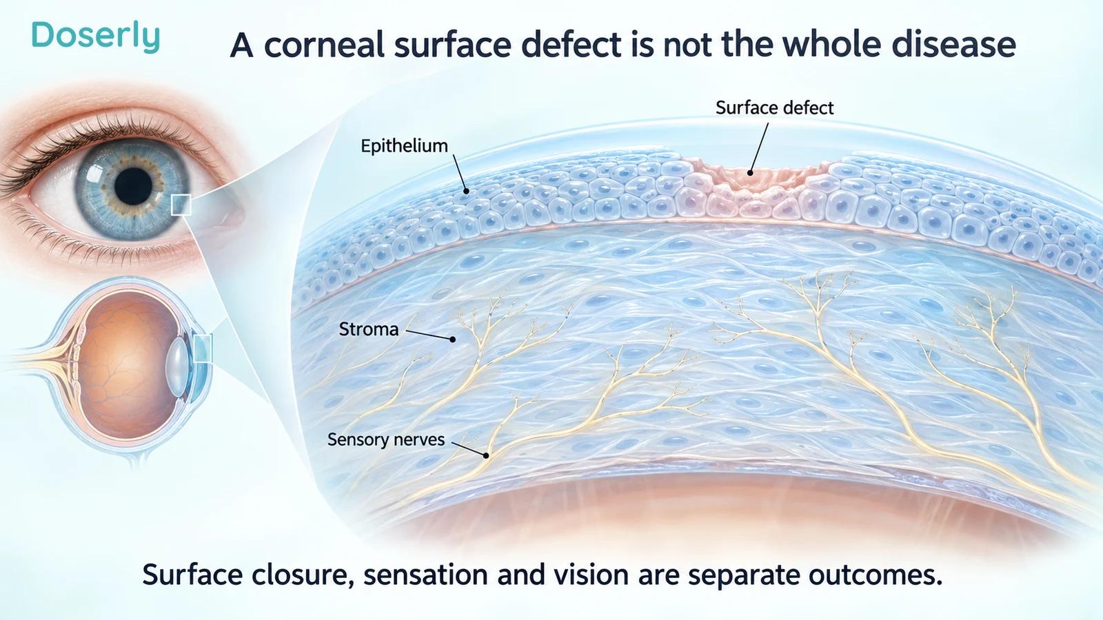
The outer layer of the cornea is the epithelium. Beneath it is the stroma, which contributes to the cornea's structure and transparency. Surface problems, deeper injury and scarring can affect vision differently. Corneal symptoms can include pain, redness, blurred vision and sensitivity to light; eye injury and sudden visual problems deserve prompt assessment. National Eye Institute corneal conditions.
Neurotrophic keratopathy involves reduced nerve support and sensation in the cornea. A defect can persist because normal maintenance and protective responses are impaired. Reduced sensation also means the amount of discomfort may not reflect how serious the surface damage is. A comfortable eye is not necessarily a healed eye.
SEER-1 enrolled adults with stage 2 or 3 neurotrophic keratopathy, persistent epithelial defects and reduced corneal sensitivity. These were not simply people with occasional dryness after screen use. Participants had a defect that had failed conventional nonsurgical treatment, and the study excluded active ocular infection. Trial eligibility and methods.
That selection matters for both benefit and safety. Someone with an infected ulcer, a new abrasion or a different cause of vision loss needs an assessment directed at that problem. Research in a selected group with neurotrophic disease cannot identify the right treatment for every painful or blurry eye.
What was the SEER-1 treatment comparison?
The trial compared 0.1% RGN-259 ophthalmic solution with its vehicle, the matching preparation without the active peptide. Ten participants received RGN-259 and eight received placebo. Treatment lasted four weeks, with follow-up visits extending to day 43. Participants and investigators were masked to assignment.
The original plan called for 46 participants. The paper says recruitment difficulties led to early termination after 18 completed the study; the registry lists termination with “Business decision” as its recorded reason. Both records describe a much smaller completed study than intended. Publication and SEER-1 registry.
The groups also differed at baseline. Placebo participants were older on average and had larger defects; the active-treatment group reported more discomfort. Randomization helps protect against systematic selection, but in a sample this small it cannot guarantee close balance. These differences complicate interpretation of subsequent changes.
A trial's “phase 3” label describes its development stage, not the amount or certainty of its evidence. Eighteen participants still produce imprecise estimates. The study remains informative because it used a control group and defined measurements, but its size is central to what can be concluded.
Surface closure: the main outcome
The primary endpoint was the proportion of participants whose persistent epithelial defect had completely healed at day 29, assessed using corneal fluorescein staining. Dye helps clinicians identify an abnormal surface. Here it was used to decide whether the defect had closed, rather than simply whether a regional staining score had improved.
At that visit, six of ten participants receiving RGN-259 and one of eight receiving placebo met the closure endpoint. Those proportions are 60% and 12.5%, a difference of 47.5 percentage points. The prespecified Fisher's exact test gave P=0.0656, which did not meet the study's conventional 0.05 significance threshold. Primary outcome, Table 2.
The observed difference is large enough to justify interest. It is also based on very few people: one participant changes the treated percentage by ten points and the placebo percentage by 12.5 points. That sensitivity is a practical way to understand why a striking percentage can remain uncertain.
The paper presents an alternative chi-square analysis with P=0.0400, but identifies it as ad hoc and acknowledges that its large-sample assumption is violated. The main comparison should retain its original interpretation. Selecting the more favorable statistical test would make the result appear more secure than the planned analysis supports.
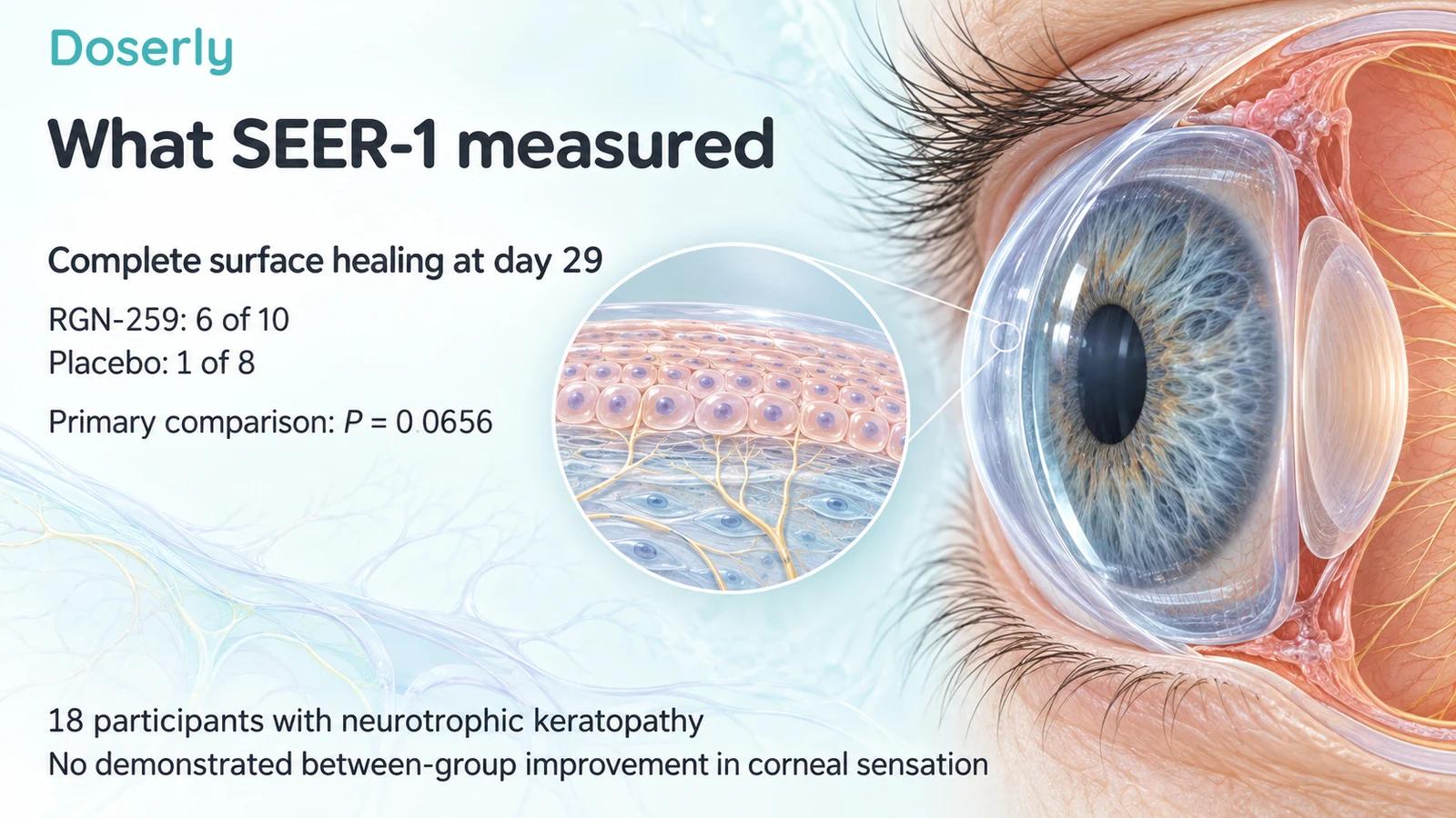
Image sources: Source
Did the surface stay healed?
Healing at one visit and durable healing are different outcomes. SEER-1 assessed closure again after treatment ended. At day 43, Table 2 reports complete healing in five treated participants and none receiving placebo, with P=0.0359. This was a later assessment, not the primary endpoint.
The report contains inconsistencies between some narrative descriptions and the visit-by-visit table. For example, the narrative describes six treated participants as healed at both days 29 and 36, while Table 2 lists six at day 29 and four at day 36. That is a reason to preserve the verified main result and avoid turning later observations into a confident claim that every healed defect remained closed. Table 2 and results text.
Two weeks of post-treatment observation is also a short period for a condition that can recur. It cannot establish whether someone remains free of defects over a year, avoids surgery or retains vision long term. Those are valuable questions for a larger study with sustained follow-up.
Comfort improved on some assessments, but that is a separate benefit
The trial asked about overall discomfort, foreign-body sensation, dryness, burning and sensitivity to light. Several comparisons favored RGN-259, including discomfort at selected visits and foreign-body sensation. The active group started with more discomfort, so the authors also presented analyses accounting for baseline values.
These measurements matter because treatment should ideally help people feel and function better as well as change examination findings. But symptom scores were secondary outcomes, assessed repeatedly in a small sample. The results should be described by symptom and timing rather than collapsed into a general statement that every symptom improved. Symptom analyses, Table 4.
For an individual patient, symptom relief and wound closure may both be meaningful. The clinician still needs to follow the surface directly, particularly when sensation is reduced. A symptom diary can describe comfort between visits, while the examination answers whether the defect is closing. Neither replaces the other.
Did thymosin beta-4 restore corneal nerves or vision?
The study measured corneal sensitivity with a Cochet-Bonnet aesthesiometer, an instrument used to assess the response to gentle contact. The report found no difference in corneal sensation between treatment groups. It also described negligible differences in mean visual-acuity change.
The authors discussed nerve repair as a possibility based on other experimental work, but explicitly stated that it was not known whether RGN-259 repaired nerve damage in neurotrophic keratopathy. They called for additional investigation, including imaging that could examine nerve changes. Discussion and safety assessments.
Surface closure therefore cannot be presented as proof that nerves regenerated. Likewise, a closed epithelial defect does not establish that scar tissue cleared or vision returned to normal. The optical quality of the cornea and other eye conditions can influence sight after the surface closes.
This distinction is useful when a proposed benefit sounds broad. “Heals the cornea” may refer only to the epithelial endpoint. Asking whether sensation, scarring and visual acuity were measured helps clarify what remains unknown without minimizing the importance of protecting the surface.
How does this differ from the dry-eye studies?
In a separate 72-person dry-eye trial, the main measurements were discomfort and lower-cornea staining scores at day 29. Neither showed a significant treatment advantage. Other regional staining and challenge-related analyses were more favorable. Dry-eye trial.
That trial did not ask how many persistent neurotrophic defects completely closed. A staining score describes the extent or intensity of a finding; a complete-closure endpoint asks whether a defined lesion has resolved. Both may use fluorescein, but sharing an examination tool does not make them the same outcome.
The populations also differ. Dry eye and neurotrophic keratopathy can coexist, but they are not interchangeable diagnoses. Combining favorable findings from both without naming the condition can make the evidence seem broader than it is. Doserly's human-research section separates the clinical settings.
What about established treatment and safety?
Cenegermin, marketed as Oxervate, is an approved ophthalmic treatment for neurotrophic keratitis. Its prescribing information describes separate controlled trials using eight weeks of treatment and a specified corneal-healing definition. That is evidence for a different drug, not a direct comparison with RGN-259. Oxervate prescribing information.
Comparing percentages across unrelated trials cannot establish that one treatment heals faster, is safer or works better. Participants, baseline severity, treatment duration and assessment methods can differ. SEER-1 did not randomize patients between RGN-259 and cenegermin, so it cannot answer which is preferable.
SEER-1 recorded 16 adverse events across seven people, with one event considered treatment-related. One serious event was considered unrelated to treatment, and no participant withdrew because of an adverse event. These details are more informative than a general label of “safe.” Eighteen participants cannot rule out uncommon harms. Safety results.
What to ask at an eye appointment
Ask which layer or function is affected and what the next visit will measure. Is the goal complete closure, preventing further damage, improving comfort, restoring sensation or improving vision? These goals may require different assessments and different treatments.
If thymosin beta-4 is discussed, ask how the proposal relates to the studied sterile ophthalmic formulation and your diagnosis. The trial does not support putting an untested research preparation into the eye, nor does it establish that an injected product reproduces the eye-drop findings.
The most useful interpretation of SEER-1 is specific: it provides a small randomized signal for epithelial closure, with a missed primary significance threshold and important uncertainty. It does not establish nerve regeneration, restored vision or lasting protection from recurrence. Keeping each outcome visible makes the research more useful for a real treatment conversation.
Sources
- SEER-1 primary publication, 2022.
- SEER-1 registry, NCT02600429.
- Dry-eye controlled-environment trial, 2015.
- Oxervate prescribing information.
- National Eye Institute: corneal conditions.