
Peptides
October 16, 2026
The 2025 Epitalon cell paper: longer telomeres are not longer life
On this page
The 2025 Epitalon paper asks a molecular question: what happens to telomere-related measurements when particular human cell lines are exposed to a peptide in the laboratory? It does not test whether people live longer, develop fewer diseases or retain independence after taking Epitalon.
That boundary is especially important because the paper includes both normal and breast-cancer cell models. A mechanism that helps maintain chromosome ends is not automatically a desirable intervention in every cell. The publication also has a correction replacing its figures, with details that deserve more attention than the headline usually receives.
The Epithalon guide covers the broader compound and evidence landscape. Here, the task is narrower: read this cell study carefully, understand what its measurements mean, and identify the steps missing between a laboratory signal and a clinical longevity claim.
Start with the molecule and the experimental setting
Epitalon, also spelled Epithalon, is the synthetic four-amino-acid peptide Ala-Glu-Asp-Gly, abbreviated AEDG. It should not be confused with epithalamin, a pineal-gland polypeptide extract. FDA's scientific review explicitly treats them as different substances. Older observations involving the extract cannot simply be assigned to the synthetic peptide. FDA review.
This is an identity issue before it is an efficacy issue. If a source uses a related preparation, its findings may help explain the history of a hypothesis, but they do not establish what the preparation in a new experiment does. A product name, a family resemblance and a shared research tradition are not evidence of equivalence.
The 2025 study by Al-Dulaimi and colleagues used 21NT and BT474 breast-cancer cell lines, IBR.3 fibroblasts and HMEC epithelial cells. The cancer-cell experiments generally involved four-day exposure, while the normal-cell experiments included three weeks. Researchers assessed telomere length, hTERT expression, telomerase activity and markers of alternative lengthening of telomeres, or ALT. These were cultured cells, not participants receiving treatment. Primary paper.
“Human cells” describes their origin. It does not mean a clinical trial was performed. In culture, cells are exposed directly in a controlled medium; the experiment does not establish absorption, distribution, clearance or exposure in a person's tissues.
Telomeres are chromosome structures, not a lifespan readout
Telomeres help protect chromosome ends. Telomerase can add telomeric repeats, while ALT is a different maintenance pathway associated with DNA-repair processes. These mechanisms are biologically important, including in cancer cells that maintain their capacity to proliferate. A large analysis across 31 cancer types examined telomere length alongside TERT and other genomic alterations, illustrating how closely maintenance biology depends on cellular context. Primary cancer-genomics study.
That makes the phrase “longer telomeres” incomplete on its own. Which cells were measured? What was their starting state? What mechanism changed? Was function preserved, and were unwanted effects assessed? The same numerical direction can have different implications in different tissues.
It is also useful to separate an average measurement from a universal claim. A result from a sampled population of cells cannot establish that every chromosome end changed equally. Nor does it reveal everything about the cells' other properties. A single measurement can be informative without serving as a complete description of biological aging.
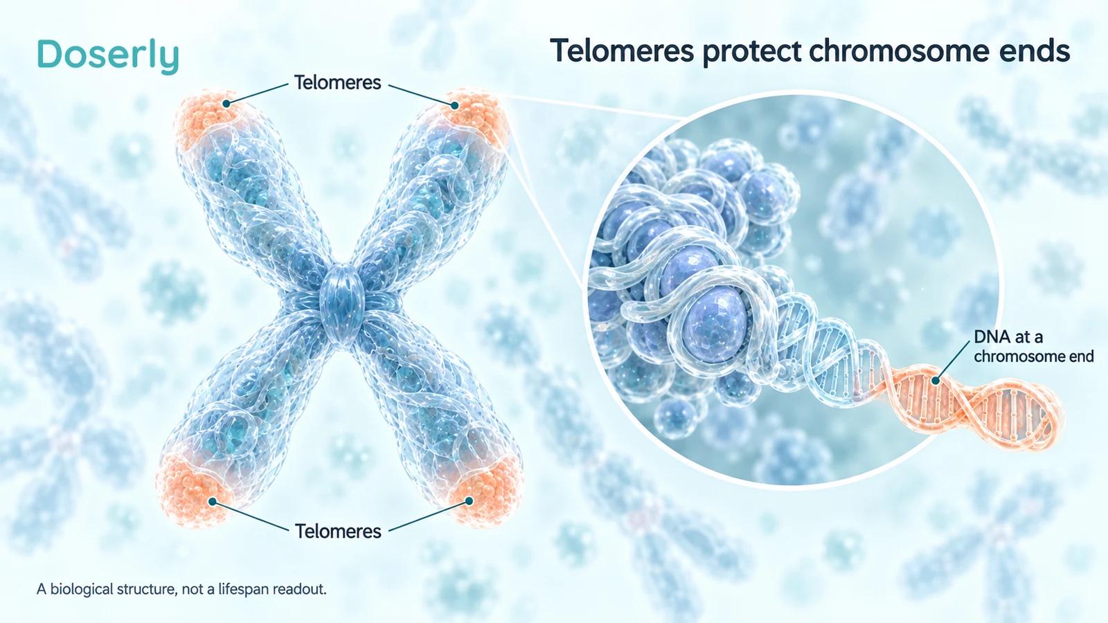
Telomeres are structures at chromosome ends. Their measurement describes a biological feature; it does not directly measure a person's remaining lifespan.
Read the correction with the paper
The correction was published online on November 15, 2025. It states that the wrong versions of Figures 1, 2 and 3 appeared, provides replacements, and says the original article was corrected. It is part of the same publication record, not a second experiment confirming the first. Publisher correction.
The corrected figures also expose reasons to avoid an unqualified summary. Figure 1C labels the IBR.3 telomere comparison “ns,” while the main text describes significant increases in both normal cell types. Figure 1D is identified as HMEC in the legend but retains an IBR.3 label beneath the panel. Figure 2G marks the HMEC hTERT comparison “ns.” Those details complicate a blanket statement that every normal-cell measurement significantly increased.
The appropriate response is to preserve the discrepancy. We should not silently choose whichever version makes the strongest story, estimate an effect from bar heights, or treat an unresolved label as settled. The broad authors' interpretation and the individual displayed comparisons are different levels of description.
A correction is not, by itself, evidence that the entire study is invalid. It is equally not a reason to ignore the corrected record. For precise quantitative claims, the text, panel identity, statistical comparison and underlying data need to agree. This article therefore avoids percentage gains and does not translate the plots into years of life.
Expression, enzyme activity and ALT answer different questions
The authors interpreted their findings as telomerase-related effects in normal cells and an ALT-associated pattern in the cancer lines. In the cancer models, increased hTERT expression did not coincide with a significant increase in telomerase activity. That distinction is central: expression of a component and measured enzyme function are not the same outcome. The corrected figures warrant the additional caution described above. Study and methods.
A useful analogy is to distinguish an instruction from the work performed. Measuring a molecular message may show that a regulatory response occurred, but it cannot replace a functional assay. Even a functional assay still needs to be connected to the outcome the reader cares about.
ALT has its own measurement challenges. A published C-circle assay protocol explains that C-rich extrachromosomal DNA circles can be used to assess ALT-associated activity, with controls and additional methods helping classify telomere maintenance. The assay is a research measurement, not a clinical longevity test. Primary methods paper.
This is why multiple measurements can strengthen an investigation without becoming interchangeable. A telomere-length result concerns the measured DNA feature. An expression result concerns a molecular transcript. An enzyme assay concerns activity under assay conditions. A C-circle result concerns an ALT-associated marker. The interpretation should name the result actually obtained.
Earlier Epitalon studies provide context, not a human endpoint
A 2003 paper reported induction of telomerase-related activity and telomere elongation in telomerase-negative human fetal fibroblast culture. It supplied an early mechanistic basis for the hypothesis. Its experimental setting remained a cell culture, even though the abstract discussed the possibility of broader lifespan effects. 2003 primary report.
A 2004 report examined proliferative capacity in fetal fibroblasts. Its abstract describes untreated cells losing proliferative potential at passage 34, while treated cells reached passage 44 and continued dividing. That is a finding about a cultured cell population. It is not an observation that a person's life increased by a corresponding fraction. 2004 primary report.
The distinction matters because “cellular lifespan” can sound like a smaller version of human lifespan. They are different endpoints. Continued division under culture conditions does not establish preserved cognition, mobility, organ function or survival in an organism. Those outcomes involve more than the ability of one cell population to keep proliferating.
The older studies help explain why the 2025 work was worth doing. They cannot fill in outcomes that the later experiment did not measure. Several papers addressing a related mechanism can build a research rationale without adding up to a clinical efficacy trial.
Human donor cells can still produce mixed laboratory results
A 2019 study used stimulated blood lymphocytes from eleven men, including five younger and six middle-aged donors. After AEDG exposure, significant telomere-length changes occurred in seven donors' samples: increases in five and decreases in two. The authors discussed a possible tendency toward normalization relative to starting length. Primary lymphocyte study.
That proposed interpretation should remain attached to its small experimental setting. It does not establish that the peptide detects a person's ideal telomere length and restores it. The cells were stimulated and exposed outside the body; the donors were not shown to obtain a clinical benefit.
This study is useful precisely because its results resist a uniform “always lengthens” story. A direction of change can differ across samples. The baseline measurement, donor characteristics, cell preparation and analysis all matter when interpreting the response. Turning selected positive samples into a universal effect would discard part of the evidence.
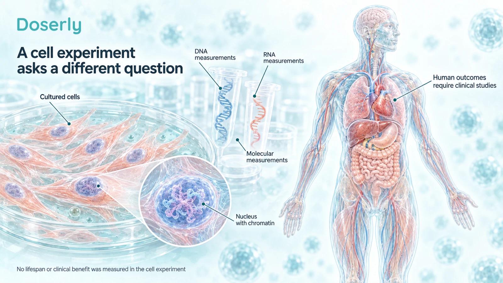
Culture experiments can investigate molecular responses. Human benefit and long-term safety require separate studies; the illustration does not imply that the laboratory findings have already transferred to people.
Why the cancer-cell findings belong in the main discussion
Cancer models are not incidental decoration in this paper. They test the intervention in cells with different biological behavior. Omitting them would make the study look more straightforwardly reassuring than it is.
Broader human genetic research also argues against treating longer telomeres as an unqualified health goal. A Mendelian-randomization analysis found associations consistent with increased risks for several cancers and reduced risks for some non-neoplastic diseases with genetically longer telomeres. Such inference depends on the method's assumptions. It does not estimate the effects or risks of taking Epitalon. Primary genetic study.
The useful conclusion is a question for development: which cells are affected, for how long, and with what consequences? The evidence does not justify claiming that this peptide causes cancer in people. It also does not justify claiming that telomere extension makes a regimen safe or protective.
Safety cannot be inferred by choosing the preferred mechanism. An intervention intended for repeated use would need evidence about unwanted effects as well as the desired molecular response. A short experiment that measures a selected pathway cannot establish that long-term balance.
A culture concentration is not a human protocol
The conditions that produce a response in a dish help another laboratory understand the experiment. They are not a dosing schedule for a person. Matching a culture concentration to a body-weight calculation would skip the evidence needed to connect administration with tissue exposure.
A meaningful development program would identify the administered formulation, characterize exposure, establish suitable safety monitoring and test an explicitly defined benefit. The relevant route and duration would need their own support. None can be selected simply by reading the axis of a laboratory figure.
The same boundary applies to product quality. A correctly identified material can still lack evidence of clinical benefit. Conversely, a paper studying a peptide does not verify the identity or quality of a separately marketed preparation. Those are distinct questions that require distinct evidence.
What would change the practical conclusion?
Independent replication could clarify the cell findings, including the discrepancies between text and corrected panels. Stronger reporting would make the relevant comparisons, variability and data accessible enough to evaluate without guessing. Reproducing a molecular effect would be progress, while remaining a preclinical result.
A clinical claim would require clinical outcomes. A trial might ask whether a defined intervention improves a particular function or reduces a particular disease outcome. It would need an appropriate comparison, sufficient follow-up and transparent reporting of adverse events. A longevity claim would need evidence matching that longer and broader promise.
Personal tracking cannot replace those comparisons. A change in energy, sleep or a commercial biomarker after several simultaneous lifestyle changes does not identify which exposure caused it. Records can help organize a discussion, but they do not validate a telomere-based treatment target.
The 2025 paper is worth reading as a study of cell biology, with its correction beside it. Its contribution is a set of model-specific observations and questions. Longer human life remains a separate proposition that these experiments did not test.
Sources
- Al-Dulaimi et al., 2025: Epitalon cell study.
- Al-Dulaimi et al., 2025: correction replacing Figures 1–3.
- Khavinson et al., 2003: fibroblast telomerase and telomeres.
- Khavinson et al., 2004: fibroblast proliferative potential.
- Khavinson et al., 2019: stimulated donor lymphocytes.
- C-circle measurement protocol, 2023.
- Barthel et al., 2017: telomeres across cancer types.
- Telomeres Mendelian Randomization Collaboration, 2017.
- FDA: Epitalon scientific review, 2026.