
Peptides
October 16, 2026
TB-500 and actin: what the cell biology can actually explain
On this page
Cells need to change shape and move when tissue responds to injury. Actin, a protein that can assemble into filaments, helps make those movements possible. Thymosin beta-4 participates in regulating actin, which is one reason the peptide appears in research on wound repair and muscle regeneration.
TB-500 is commonly discussed alongside that biology. The connection is real, but the usual shorthand can be misleading. “Binds actin” does not mean “builds muscle,” and a fragment containing an actin-related sequence does not automatically reproduce the behavior of the full peptide.
The useful question is how these molecular findings relate to something a person would notice: a wound closing, strength returning or an injured limb functioning better. To answer it, we need to keep the full peptide, the fragment, the experimental setting and the measured outcome distinct.
Actin is dynamic, not a fixed scaffold
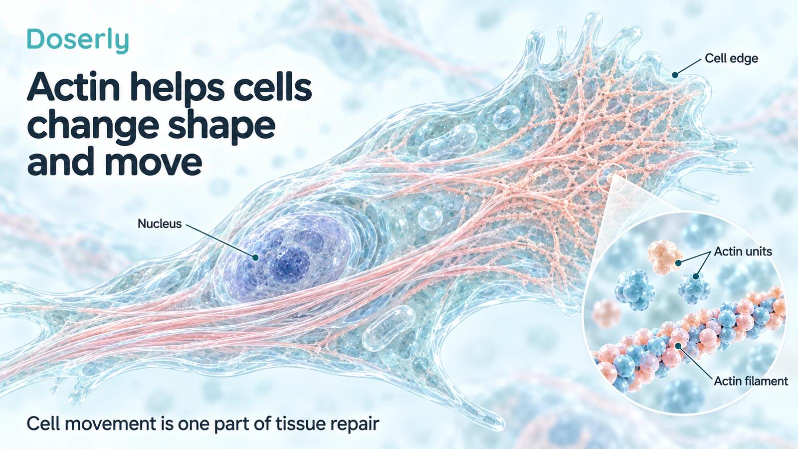
Actin exists as individual units, often called globular or G-actin, and as assembled filaments, called F-actin. Cells regulate the supply and arrangement of both. Assembly and disassembly help a cell reshape its edge, organize internal structures and respond to its surroundings.
Thymosin beta-4 and another protein, profilin, help regulate the pool of actin units. Structural research shows that thymosin beta-4 can bind an actin unit in a way that prevents it from joining a filament. Exchange with profilin is part of the system that makes actin available for assembly. Primary structural study, 2014.
This may sound surprising if you've heard that thymosin beta-4 simply “increases actin.” Its role is more about controlling availability and interactions than adding building material. A functioning cell needs organized turnover, not the maximum possible amount of filament at every moment.
The distinction also separates cell movement from muscle contraction. Both involve actin, but observing an effect on a cultured cell's movement does not show that a person's muscle will generate more force. The structures, surrounding signals and outcome being measured differ.
Which peptide does the actin research concern?
Full-length thymosin beta-4 contains 43 amino acids. The TB-500 substance evaluated in FDA's 2026 briefing is an N-acetylated seven-amino-acid fragment, Ac-LKKTETQ. It corresponds to a region within the longer peptide. FDA TB-500 briefing.
That shared region explains the scientific interest. It does not establish that the fragment has the same binding behavior, distribution or clinical effects. The rest of the molecule can contribute to its interactions, and a modification at an end can matter as well.
A primary study that altered individual amino acids in full-length thymosin beta-4 found that residues distributed across the peptide contributed to actin binding. Changes outside the short LKKTETQ region could substantially weaken binding. Mutational analysis, 2008.
This is a specific reason to be cautious with the phrase “the active part.” A region can be important without being sufficient for all of the larger molecule's behavior. Our TB-500 identity guide explains the chemical distinctions without treating the names as interchangeable.
Why more thymosin beta-4 does not always mean more movement
A 2014 laboratory study examined interactions among thymosin beta-4, cofilin and the Arp2/3 complex, proteins involved in actin dynamics. In HeLa cells, increasing intracellular thymosin beta-4 reduced actin filament bundles and cell migration under the tested conditions. Reducing its expression produced different effects. Primary cell study.
HeLa cells are a laboratory cell line, not injured human muscle. The result should not be read as proof that thymosin beta-4 would slow a person's healing. Its importance is that it contradicts a universal one-direction story. The effect depends on the cellular setting and the balance of interacting proteins.
Changing the amount of a peptide inside a cell is also different from injecting a preparation outside cells. Researchers would need to establish whether an administered product reaches the relevant compartment and at what exposure. A mechanism demonstrated by changing gene expression is not automatically recreated by an injection.
For a recovery claim, the question is therefore more detailed than whether actin is involved. It is whether the particular product changes the relevant process, in the relevant tissue, in a way that improves the patient's outcome.
What the muscle-injury study actually examined
A study published in 2011 investigated thymosin beta-4 during skeletal muscle regeneration in mice. Researchers found increased expression in regenerating muscle and inflammatory cells after injury. They also examined how muscle precursor cells, called myoblasts, responded in laboratory migration experiments. Primary muscle-injury study.
The work included thymosin beta-4 and a modified, oxidized form. The researchers reported chemotactic effects, meaning movement of cells toward a chemical signal. Experiments included a mouse myoblast cell line and primary cells derived from adult mouse muscle satellite cells.
That supports a role for thymosin beta-4-related signaling in the response to injury. It does not show that giving the TB-500 fragment to people speeds return to sport. Increased production of the body's own peptide after an injury is not proof that administering more is beneficial, and the oxidized form should not be treated as identical to every other preparation.
The distinction between expression and treatment is especially important here. Measuring what the body produces tells us about the injury response. A treatment study asks whether changing that response from outside improves recovery. The second question does not answer itself once the first is observed.
What the short-fragment wound study contributes
A 2003 study examined dermal wound repair in diabetic and aged mice. Full-length thymosin beta-4 showed activity in these models. A synthetic LKKTETQ fragment also promoted repair in aged animals under the experimental conditions. Primary mouse wound study.
That is useful evidence that the short sequence can have biological activity. But the fragment in this study was not automatically the N-acetylated Ac-LKKTETQ substance represented by TB-500 in later analytical work. The result concerned mouse skin wounds, not a measured human muscle or tendon benefit.
It would be an overcorrection to say that all fragment biology is invented. It would be an overstatement to call the animal result a validated human recovery protocol. The accurate description sits between those claims and remains specific about the preparation, species and outcome.
Even within wound repair, changes in cell migration, collagen deposition and closure are different observations. They can help explain an experiment, but none should be silently replaced with a claim about the strength of a healed human tissue.
Newer TB-500 research raises a metabolite question
A 2024 analytical study examined TB-500 and its breakdown products in laboratory systems and in rats. It also compared their activity in a fibroblast wound-healing assay. In that assay, the metabolite Ac-LKKTE, rather than the parent compound, was the one reported to show significant activity against the control. Primary metabolism and cell-assay study.
A metabolite is a substance produced when another substance is transformed or broken down. This finding suggests that researchers should distinguish exposure to the parent peptide from exposure to its products. It does not establish that a metabolite heals human wounds or justify changing a dosing schedule.
The experiment also included human serum in vitro. That means serum was used outside the body as an experimental environment; it does not mean human participants received TB-500. Rat findings likewise remain rat findings.
For the clinical question, the missing steps include whether the relevant metabolite forms in people, where it reaches, how long it remains and whether its presence improves a meaningful outcome. The study offers a more precise hypothesis, not a completed answer.
Why a cell-culture gap is not a healed injury
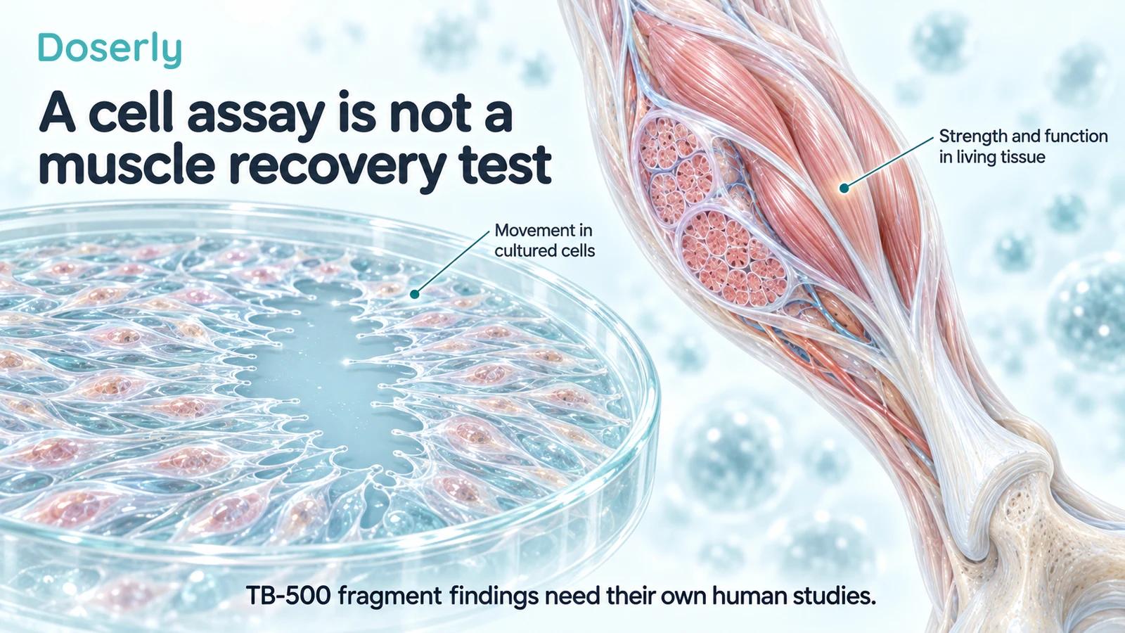
In a laboratory migration or wound-closure assay, researchers can observe cells moving into an open area. That makes the experiment useful for studying cell behavior under controlled conditions. It does not reproduce a full wound with blood supply, immune responses, connective-tissue organization and mechanical loading.
The measured result can also depend on the assay's design. Cell movement, cell survival and cell proliferation may contribute differently to the appearance of a closing gap. Researchers need methods that help distinguish those processes when making a mechanistic claim.
A tendon or muscle injury adds further questions. Can the repaired tissue tolerate load? Does pain improve during meaningful activity? Does function remain better after treatment ends? A culture experiment cannot measure those patient outcomes, even when its biological result is reproducible.
This is why the microscope image should be described as a cell result. Calling it tissue regeneration can imply a much broader conclusion than the experiment supports. The more carefully the assay is named, the easier it is to understand what the next study should do.
What would translate the mechanism into a useful treatment?
A development program would need a well-characterized product and measurements of its behavior after administration. The fragment, any metabolites and the target tissue would need attention. Without that information, a laboratory concentration cannot be assumed to correspond to a practical human exposure.
Researchers would then need a defined clinical problem. A chronic skin wound, an acute muscle tear and a surgically repaired tendon require different outcomes and background care. An effect in one condition would not automatically become a general recovery benefit.
For a muscle injury, a useful study might measure strength and return to meaningful activity alongside symptoms and safety. For a wound, durable closure and recurrence could be important. These are examples of research questions, not evidence that TB-500 has already achieved those results.
FDA's review did not identify human clinical evidence for the specific TB-500 substances it evaluated. That leaves the translation question open despite the interesting actin and cell-migration literature. FDA briefing.
What to take into a recovery conversation
If a treatment is explained to you as “activating actin,” ask what that phrase means in the cited experiment. Was the work done with full-length thymosin beta-4, an unacetylated fragment, N-acetylated TB-500 or a metabolite? Was it a cell assay, an animal injury model or a human treatment trial?
Then connect the study to your goal. A person hoping to regain shoulder strength needs different evidence from someone hoping for a skin wound to close. A plausible mechanism can justify research, but it should not be used to promise a result that was never measured.
The TB-500 human-evidence guide places these findings alongside the available clinical work on related preparations. Actin biology helps explain why scientists are interested. It does not yet provide a reliable human recovery benefit, injection schedule or safety estimate for the fragment.
Explore the research
- Thymosin beta-4/profilin structural study, 2014: regulation of actin availability and filament assembly.
- Actin-binding mutational analysis, 2008: contributions from multiple regions of the full peptide.
- Actin dynamics and HeLa migration, 2014: context-dependent effects of intracellular thymosin beta-4.
- Mouse muscle-injury study, 2011: endogenous expression and myoblast chemotaxis.
- Mouse wound and fragment study, 2003: full peptide and LKKTETQ in animal models.
- TB-500 metabolism and cell assays, 2024: parent versus metabolite activity.
- FDA TB-500 briefing, 2026: fragment identity and clinical-evidence gaps.