
Red Light Therapy
October 17, 2026
Red light therapy wavelengths and power: a complete guide to the evidence
On this page
A red light device can come with a surprisingly long list of numbers: 630, 660, 810, 830, 850 or 1064 nanometers. The explanation often sounds reassuringly precise. One wavelength is said to treat skin, another to reach muscles, and another to stimulate the brain. It can make choosing a device feel like matching a number to a body part.
The science is more interesting, and less tidy. Wavelength affects how light travels through tissue and which molecules absorb it. But a wavelength does not come with a fixed depth, a guaranteed effect or a complete treatment plan attached.
Red and near-infrared light have different optical properties, but there is no universal best wavelength. The relevant question is whether a particular light exposure reaches the intended tissue safely and produces a useful result in people with the condition being treated. Human studies can help answer that question. A wavelength chart alone cannot.
This article explains the physics behind the common numbers, then looks at what actual human and human-tissue studies can tell us. Research available through October 17, 2026 is considered, including newer studies that measure biological responses without establishing a clinical treatment benefit.
First, what does a wavelength tell you?
Wavelength describes the spacing of an electromagnetic wave. In these device descriptions it is usually expressed in nanometers, abbreviated nm. Red light is part of the visible spectrum. Near-infrared lies beyond the red end of ordinary human vision. Both can carry energy even when the eye cannot judge the exposure.
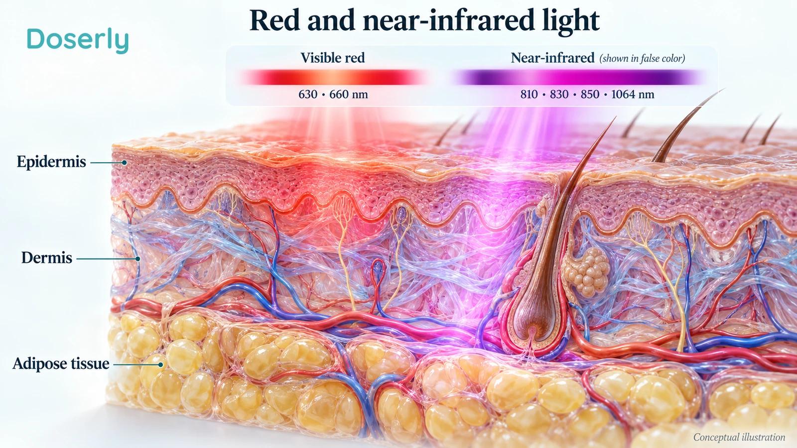
Numbers such as 630 and 660 nm describe red light. The 810, 830 and 850 nm ranges commonly used in PBM research are near-infrared, as is 1064 nm. Boundaries between named spectral bands depend on the convention being used, so the actual wavelength is more informative than a broad label such as “infrared.”
Wavelength is different from brightness, power and total exposure. A device emitting at 660 nm can deliver a small amount of light or a large amount. Two 850 nm devices can illuminate very different areas. A visible red glow may also come from only one part of a device's output while a near-infrared component remains invisible.
An LED usually emits across a band of wavelengths around a central value. A laser generally has a much narrower spectrum. That distinction can matter for describing an experiment, but it does not mean a laser is automatically a better treatment. The clinical effect depends on the entire exposure and the tissue receiving it.
Reporting standards therefore ask researchers to describe wavelength alongside power, exposure time, beam area, pulsing, treatment location and schedule. Without those details, it can be difficult to reproduce an experiment or compare it with a home device. Photomedicine reporting recommendations.
What happens when light meets skin?
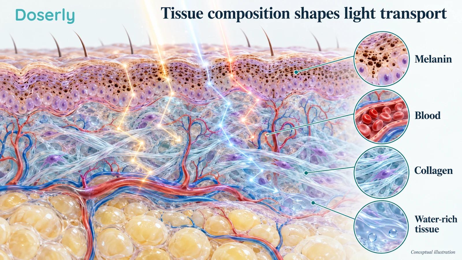
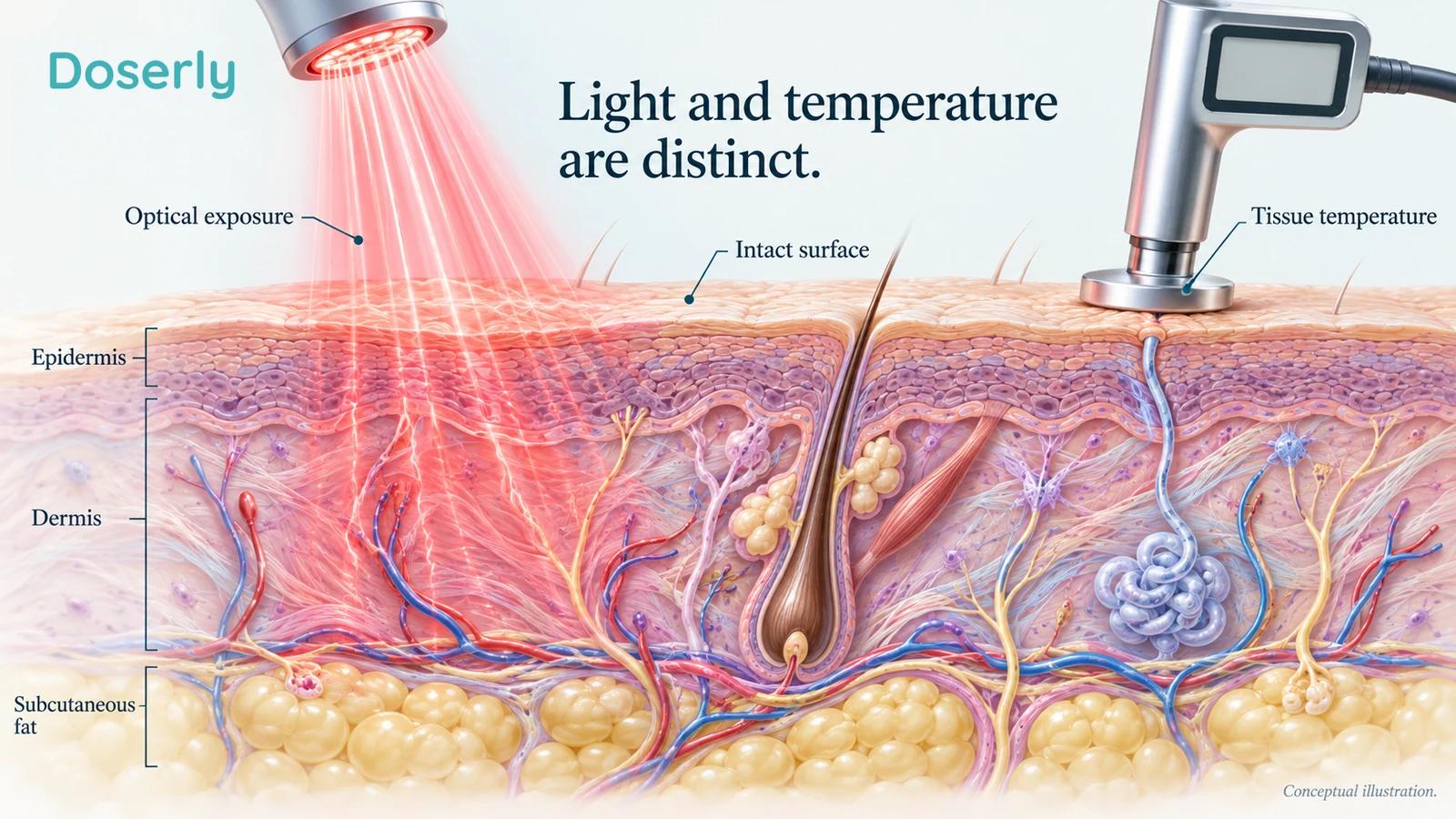
Some light is reflected at the surface. Light that enters can be scattered, changing direction, or absorbed, transferring its energy to tissue. These processes happen repeatedly in a complex mixture of cells, fibers, fluid, blood and pigment.
Absorption and scattering are different. Absorbed light is no longer continuing along its previous path as a photon. Scattered light may still travel through tissue, but its route becomes less direct. A useful picture is a network of changing paths, not a perfectly straight beam that stays intact until it reaches a predetermined depth.
The balance changes with wavelength and tissue composition. Melanin, hemoglobin, water and other tissue constituents contribute differently across the spectrum. Tissue-optics research describes these wavelength-dependent properties using measurements and models. It does not assign every person the same exposure beneath the skin. Jacques' tissue-optics review and supporting material.
This distinction helps explain why “deep penetration” is incomplete as a treatment claim. If a detector can register a small amount of light after it passes through tissue, that establishes transmission under those conditions. It does not show that the remaining exposure is sufficient to change a disease outcome. Nor does it show that increasing the output until more light is detected would be comfortable or safe.
Conversely, a superficial target does not need the greatest possible depth. If the intended effect concerns structures within skin, light that reaches far beyond them is not automatically more useful. The best optical match, if one can be established, depends on where absorption is needed and what biological response is being sought.
A layer diagram is useful for understanding this, but it should not be mistaken for a personal map. Real tissue has boundaries, curves, variable thickness and blood flow. The same named body region can contain very different optical paths in different people.
Why exact penetration charts can mislead
“Penetration depth” can mean different things in different contexts. It may refer to a calculated decline in light intensity, the point where a detector reaches its sensitivity limit, or a depth at which a researcher believes a biologically relevant exposure is possible. Those are not interchangeable definitions.
That is why a chart stating that red light reaches one fixed depth and near-infrared another should prompt a follow-up question: how was the depth defined and measured? Was the work done in a living person, excised tissue, a cadaver, an animal or a computer model? What was the input exposure, and what proportion remained?
A 2023 review of measured human skin optical properties found substantial variation both within and between publications. Measurements made outside the body were especially variable for absorption. This makes a single definitive optical value at a given wavelength difficult to defend across all settings. In-vivo and ex-vivo skin optics review.
Original tissue research also illustrates the limits of simple charts. One study measured 660 nm transmission through sites in living people and cadaveric tissue. Tissue thickness, composition and measurement conditions affected the findings. The authors did not find a skin-tone effect within their study, but that result should not be turned into a universal claim that pigmentation never matters for light delivery or skin responses. Human red-light transmission study.
For a reader comparing devices, the practical lesson is to resist a depth claim that omits its method. “Reaches muscle” can sound like proof of a muscle treatment even when the supporting experiment only measured light on the far side of a tissue sample. The measurement may be scientifically useful; the extra therapeutic conclusion still needs its own evidence.
Red light around 630 and 660 nm
Red wavelengths are commonly studied for skin-related outcomes and other targets accessible near a surface. Their visibility makes them easy to recognize, but visual familiarity should not be confused with a complete understanding of their biological effects.
A split-face clinical study published in 2007 tested 633 nm, 830 nm, their combination and sham treatment in 76 people with facial wrinkles. The investigators assessed clinical appearance and several tissue-level measures. The study is useful because it considered different light settings within one research program, rather than comparing unrelated advertisements. Its reported improvements were not a guarantee that every red or near-infrared mask would produce the same result. Lee and colleagues' controlled study.
More recent work also shows why choosing a wavelength is only the beginning. A 2025 facial rejuvenation study used a 660 nm mask and compared application schedules with sham. Its different measurement methods did not all agree about benefit. That is a reminder that specifying a familiar wavelength does not make the clinical result simple or uniform. 2025 facial rejuvenation study.
There is no reason to assume that 630 and 660 nm are identical. There is also no sound basis for declaring one universally superior because the number is higher, lower or closer to a proposed absorption peak. To make that claim, a comparison would need to control the other important variables and measure the outcome of interest.
Imagine two masks with different wavelengths, fit, output and session schedules. If one produces better results in separate studies, the difference could be due to any of those features, the participants or the study design. Wavelength may contribute, but the comparison has not isolated it. This is why a precise-looking number can create more certainty than the evidence actually provides.
Near-infrared around 810, 830 and 850 nm
Near-infrared wavelengths are often investigated when researchers want light to travel beyond the most superficial tissue layers. In some optical circumstances they are transmitted more favorably than visible red. The qualification matters: the advantage depends on the tissue, wavelength and measurement geometry.
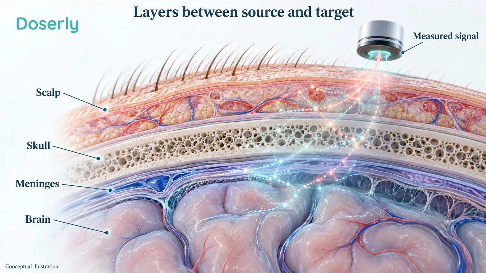
A cadaver study examined light propagation through scalp, skull and brain and compared several wavelengths within brain tissue. Under those experimental conditions, 808 nm showed less absorption and scattering than 660 or 940 nm. The work involved eight cadaver heads, not a clinical trial demonstrating benefit in living patients. The authors explicitly recognized that cadaver measurements do not reproduce every living condition. Human cadaver brain-transmission study, published figure-color correction.
It would therefore be an overreach to translate those findings into “808 nm reaches everyone's brain at a useful dose.” They provide information for further research. They do not bypass the need to establish appropriate delivery, temperature, safety, patient selection and clinical outcomes.
There are also important differences between measuring through tissue and treating it. A sensor on the far side collects whatever light reaches its location. A biological target is distributed through a volume and may respond to the pattern of absorbed energy. The sensor reading and the target's response are related questions, but they are not the same measurement.
The closeness of 810, 830 and 850 nm on a product specification does not prove their equivalence. Equally, a small difference in central wavelength does not automatically make one clinically superior. Evidence from a particular 830 nm device should be described as evidence about that intervention unless a broader equivalence has been demonstrated.
What is different about 1064 nm?
The 1064 nm wavelength is also near-infrared and appears in both experimental PBM and other kinds of medical laser use. The shared wavelength does not make those treatments equivalent. A laser used to deliberately heat a target or remove hair has a different exposure strategy from a PBM experiment.
Human physiological research has compared several near-infrared sources. In a forearm experiment, two groups of ten healthy participants were assessed with near-infrared spectroscopy during active and sham exposures. The researchers compared 800, 850 and 1064 nm lasers and an 810 nm LED, observing changes in measures related to oxygenated hemoglobin and oxidized cytochrome c oxidase. Human forearm wavelength experiment.
Those findings show that investigators can detect physiological responses under controlled conditions. They do not tell us that the wavelength producing the largest or longest signal will be the best treatment for pain, wrinkles or a neurological disorder. The participants were healthy, the target was the forearm, and the outcomes were biological measurements rather than recovery from disease.
A 2026 study examined 1064 nm stimulation in 25 healthy adults using brain electrical and magnetic recordings. It reported changes in network activity. This adds to mechanistic research, but network reorganization is not itself proof of improved everyday cognition, durable symptom relief or disease prevention. 2026 cortical dynamics study.
The important question is what a new number adds to a treatment decision. If a seller highlights 1064 nm as an upgrade, ask whether the cited evidence concerns the same target, delivery method and outcome you care about. A more unusual wavelength can be scientifically interesting without making a device a better choice for your situation.
Skin tone, tissue composition and geometry matter
Light encounters a layered biological environment, not a uniform slab. Skin pigment, blood content, tissue thickness and the arrangement of structures affect how exposure is distributed. A treatment delivered to a cheek is not optically equivalent to the same device directed at a scalp covered by hair.
A 2024 review specifically examined skin color and optical properties. It found meaningful variation in the published measurements and discussed implications for optical technologies. This is a reason to improve representation and measurement, rather than assume that one estimate applies equally to everyone. Skin color and optical properties review.
Laboratory measurements of separated epidermis, dermis and subcutaneous fat across a broad wavelength range further show why the tissue layers need to be considered individually. Their optical characteristics are useful inputs for modeling energy distribution, but a model based on sampled tissue is still an approximation of a living person. Layer-specific human tissue measurements.
Even the path used to measure light can change the interpretation. In a study involving ten patients undergoing neurosurgery, investigators examined how near-infrared measurements changed across tissue interfaces. Their findings challenged the assumption that superficial tissues make only a minor contribution to the detected signal. This was research about optical measurement, not a treatment trial. Near-infrared behavior in the adult human head.
These studies help explain why a universal calculator cannot turn a product label into a reliable dose at a deep target. They also support a more sensible standard for device claims: the closer the tested situation is to the proposed use, the more informative the evidence becomes.
Does combining wavelengths improve results?
A combination can have a plausible rationale. Different wavelengths may distribute energy differently or interact with different tissue constituents. But plausibility alone does not establish that a combination is more useful than either component on its own.
The cleanest clinical comparison would test the combination against each component, with carefully described exposure and an appropriate control. It would also address whether the combined device simply delivers more total energy or covers more tissue. Without that information, a benefit attributed to “synergy” may have another explanation.
A 2025 hair study compared 650, 1550 and 14,000 nm approaches with a control group in 68 participants. It reported hair-density changes during a treatment and maintenance period. These widely separated wavelengths do not form a simple comparison between the common red and near-infrared options, and the accessed abstract does not establish that the results isolate wavelength independently of other delivery differences. Three-wavelength hair study.
That study also shows why the word infrared needs detail. Far-infrared at 14,000 nm is not the same optical category as 810 or 850 nm. Putting all of them under one familiar marketing term hides differences that matter when interpreting tissue interaction and treatment design.
A combination study can still be useful even if it cannot isolate every component. It may tell us whether the package of treatment helped under the tested conditions. The mistake is to turn that package-level result into proof that each wavelength is essential, that adding still more wavelengths would help, or that a different combination is equivalent.
For a reader, the most useful question is therefore not how many wavelengths a product contains. It is whether the complete intervention has evidence for the intended use and whether the claimed contribution of each wavelength was actually tested.
Why an absorption peak does not prove a clinical benefit
An absorption spectrum describes how a material interacts with light across wavelengths. An action spectrum describes how a biological response varies with wavelength. The two can be related, but they answer different questions.
A molecule might absorb light in an isolated experiment without being the dominant absorber in intact tissue. Even if it participates in a response, that response may depend on cellular state, oxygen availability and other conditions. Moving from a molecular result to a treatment outcome therefore requires several additional steps.
The cytochrome c oxidase explanation often used in PBM discussions is a useful example. Researchers have proposed that interactions involving this mitochondrial enzyme and nitric oxide contribute to some effects, while other investigators have questioned how directly the available experiments establish that mechanism. Critical review of the CCO and nitric oxide evidence.
For the person choosing a treatment, there is no need to settle every mechanistic dispute first. A well-designed clinical trial can demonstrate a useful outcome even while the full explanation remains uncertain. What should be avoided is the reverse move: treating an attractive mechanism as if it already demonstrated a useful clinical outcome.
This is particularly relevant when mitochondrial language is used to connect unrelated interventions. The Doserly SS-31 guide describes a different mitochondrial research story with its own clinical context. Sharing a discussion of mitochondria does not make a peptide, a red-light mask and a near-infrared laser equivalent treatments or establish a reason to combine them.
Power starts at the wall, but treatment happens at the tissue
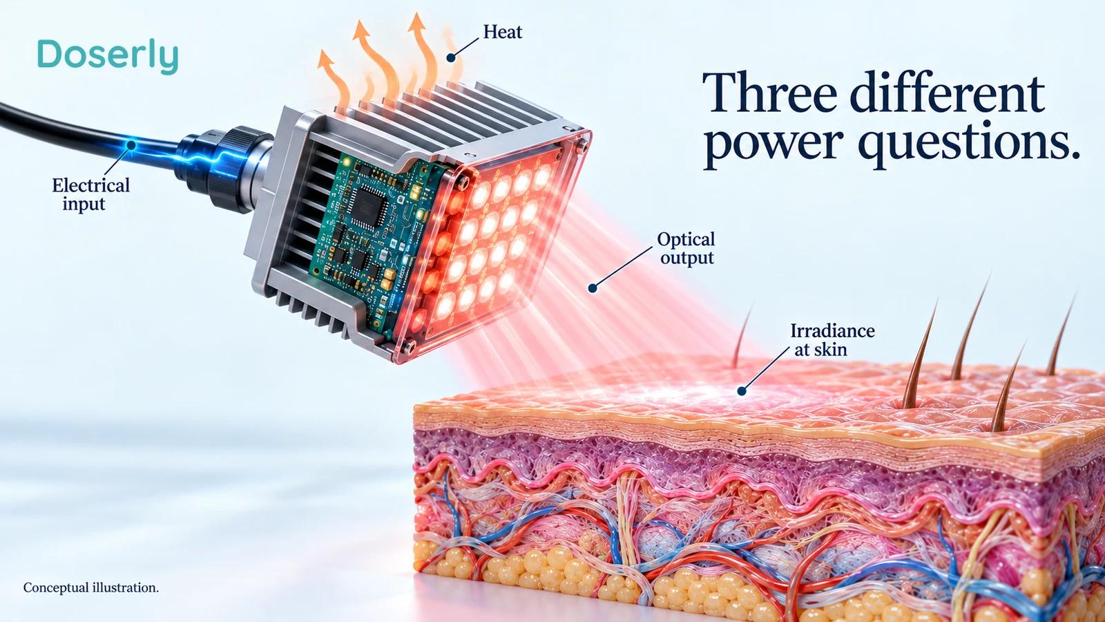
A device may carry several wattage figures, all accurate in their own context. The electricity it draws is one figure. The combined nominal ratings of its LEDs may be another. The optical power actually emitted is a third. None should be substituted for another without explanation.
Electrical input supports light generation, electronics and losses that include heat. Optical output is the radiant energy leaving the source each second. Irradiance then describes how quickly radiant energy arrives per unit area at a specified location. A large panel can have substantial total optical output while distributing it over a much larger area than a small clinical probe.
The unit relationship is straightforward: a watt is a joule per second. The NIST description of SI units supplies the basis for that calculation. It does not imply that watts are a measure of clinical effectiveness. A large electrical rating cannot establish how much useful light reaches a cheek, and a high optical output cannot establish which outcome will improve.
An informative specification names the quantity and its measurement conditions. If the page says only “power,” ask whether that means wall consumption, nominal LED capacity, measured total optical output or irradiance at a particular distance. A number without that context is hard to compare fairly.
This matters especially when switching between devices. A handheld probe, a fitted mask and a full-body panel can share a wavelength while producing different distributions of light. The exposure tested in a clinical trial belongs to that complete arrangement, including how the source was positioned and how the relevant surface was covered.
Irradiance and fluence: useful arithmetic with clear boundaries
Irradiance is commonly listed in milliwatts per square centimeter. Surface radiant exposure, often called fluence in PBM reports, is commonly listed in joules per square centimeter. One describes the delivery rate per area; the other describes the accumulated energy per area during an exposure.
For a hypothetical continuous source, 10 mW/cm² equals 0.010 W/cm². Over 400 seconds, that would deliver 4 J/cm² at the measured surface. The example is deliberately arithmetic only. It is not a recommendation for skin, hair, pain, eyes or any other target.
The calculation requires the average irradiance actually arriving at the surface during that time. It does not accept the device's electrical consumption as a substitute. Nor does it calculate energy absorbed at a particular depth. These distinctions are easy to lose when a calculator asks for just two numbers and returns a reassuringly precise answer.
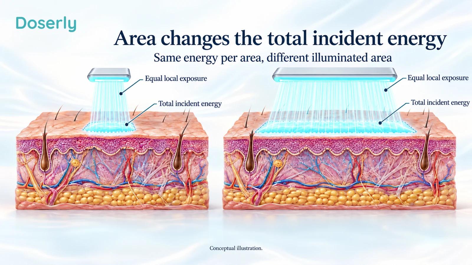
Area matters as well. Four joules per square centimeter over a uniformly exposed 10 cm² patch would correspond to 40 joules arriving at that patch. Over 100 cm², the same local radiant exposure would correspond to 400 joules. Those are different total energies despite identical exposure per unit area. The mathematical relationship says nothing about whether either example is therapeutically appropriate.
Equal energy per area also does not prove biological equivalence. Changing intensity and duration while keeping their product fixed may change temperature, signaling or the temporal pattern experienced by the tissue. Clinical interchangeability has to be demonstrated, rather than assumed from dimensional analysis.
Coverage and uniformity can matter more than the peak number
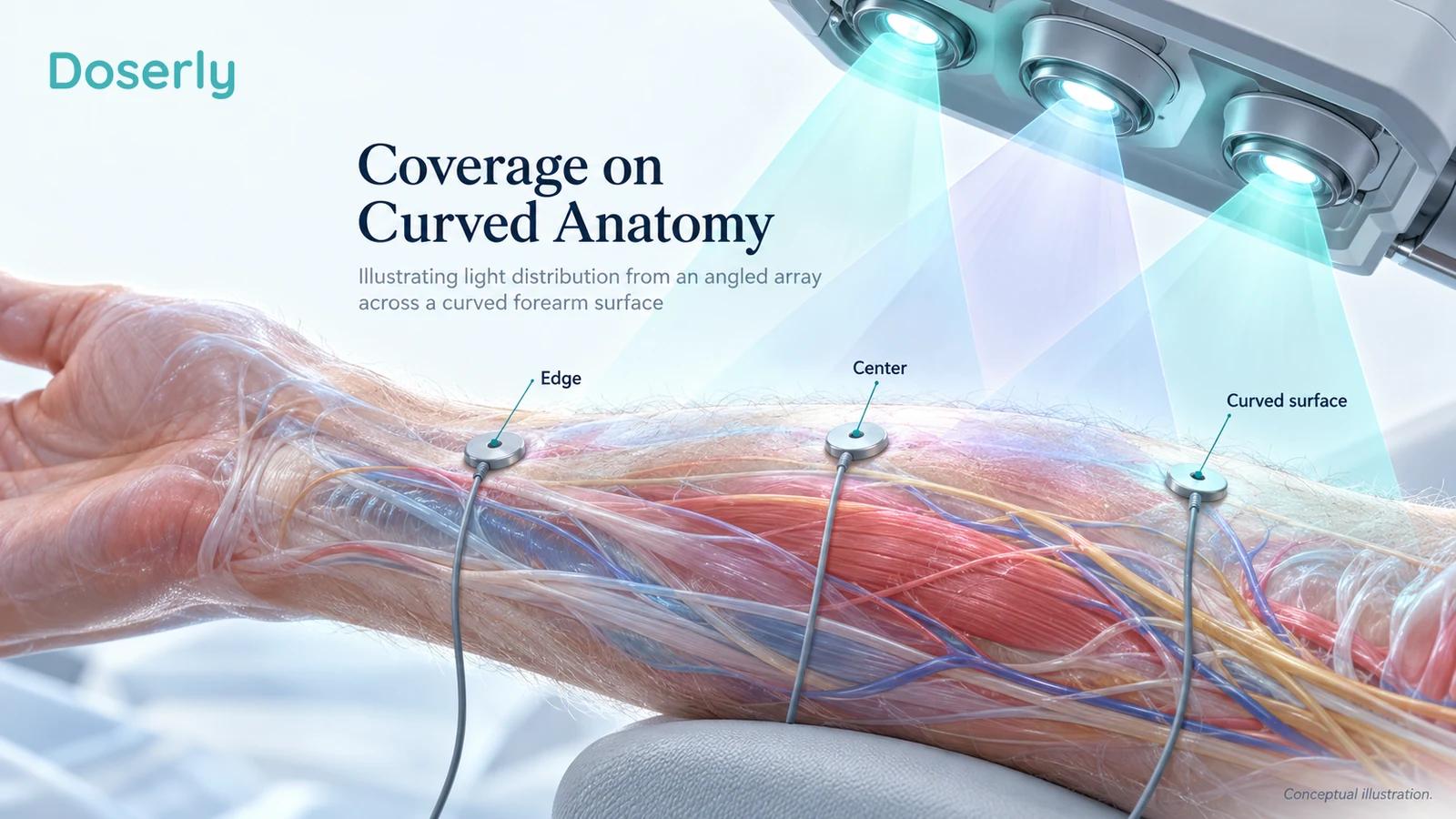
A panel is an array of emitters and optics, not a mathematical point. At close distances, different beams overlap in ways that depend on their spacing, divergence and lenses. The distribution can change as the source moves, even if the electrical setting stays unchanged.
That is why the simple inverse-square rule for an ideal point source should not be applied automatically to a nearby LED array. Actual measurements at the proposed treatment plane are more useful. The panel's central maximum, a spatial average and its weaker edge regions describe different aspects of delivery.
A good report tells you which of those was measured. It also identifies the area over which an average was calculated. Without that information, two products may appear to differ in power when their sellers have simply chosen different ways to summarize their output.
There is empirical reason to care. A 2022 characterization of 24 clinical laser devices found discrepancies between declared and measured power and beam dimensions, as well as differences across equipment. Many devices were old, so the sample cannot be used as a blanket indictment of today's consumer market. It does demonstrate that clinical ownership does not replace calibration.
A 2025 pilot examining five home-use LED devices similarly found heterogeneous optical characteristics relevant to reproducible exposure. Its tissue heating experiments used porcine tissue, not people undergoing treatment. These studies support asking for measurements; they do not identify a universally superior brand or form factor.
Curved anatomy makes a flat measurement plane only an approximation. The forehead, nose, cheek and jaw sit at different angles. Hair may intervene between a scalp device and skin. Contact pressure can change geometry and local tissue conditions. A well-designed application tries to make these factors repeatable, rather than pretending they disappear.
Measuring light requires the right detector
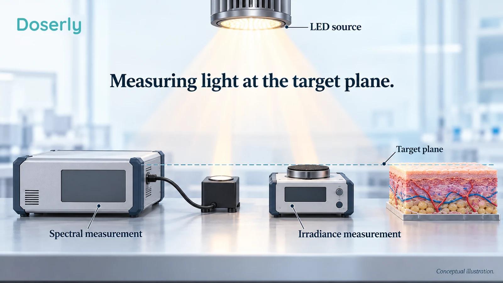
A meter is useful only if its response matches the quantity being measured. A household light meter commonly reports illuminance in lux, which is weighted to human visual sensitivity. PBM output is generally discussed in radiometric units, which describe radiant energy rather than perceived brightness. NIST's introduction to the candela explains this distinction.
Near-infrared makes the problem especially clear: a device can emit substantial radiation that the eye cannot see. A visible red glow cannot tell you the near-infrared contribution. A phone camera may detect some wavelengths, but its filters, exposure processing and sensor response do not turn a photograph into a calibrated dose measurement.
Useful optical characterization specifies the detector, wavelength calibration, measurement distance and sampled area. Spectral measurements help establish what wavelengths are present. Power and irradiance measurements answer different questions about how much radiation is delivered. No single photograph of a handheld meter settles all of them.
Consider a mixed red and near-infrared panel. If the detector responds differently to the two spectral bands, its combined reading needs appropriate interpretation. If individual channels can be switched, measurements of each channel and the combination may clarify the exposure. The point is transparency, not an expectation that every home user buy laboratory equipment.
For most readers, the practical role of measurement literacy is to judge the quality of documentation. A clear independent report is more informative than an unexplained meter display, especially when a purchasing decision depends on a small advertised difference between devices.
What to do when a specification is missing
Missing measurements should remain missing in your interpretation. If a manufacturer provides a wavelength and session duration but no usable irradiance information, you cannot reliably reconstruct the surface exposure from the number of LEDs. Counting emitters does not reveal their operating current, optical efficiency, lenses or distribution across the target.
It is reasonable to ask for the measurement report rather than reverse-engineer an answer from promotional material. The report should identify the particular model and operating mode. A test of a smaller panel or an earlier version may be informative background, but it does not automatically characterize the product being sold today.
An honest uncertainty range can be more useful than a single unsupported value. If the measurement varies across the treatment area, the variation belongs in the description. If output changes as a device warms up, the relevant exposure depends on what happens over the session, not just the first reading. These questions help distinguish reproducibility from numerical decoration.
The same standard applies to research papers. A missing technical detail does not prove that a positive result is false. It does limit how confidently someone can reproduce the intervention or transfer it to another device. That is a practical limitation worth preserving when discussing the findings with a clinician or comparing purchase options.
Pulsing changes the exposure pattern
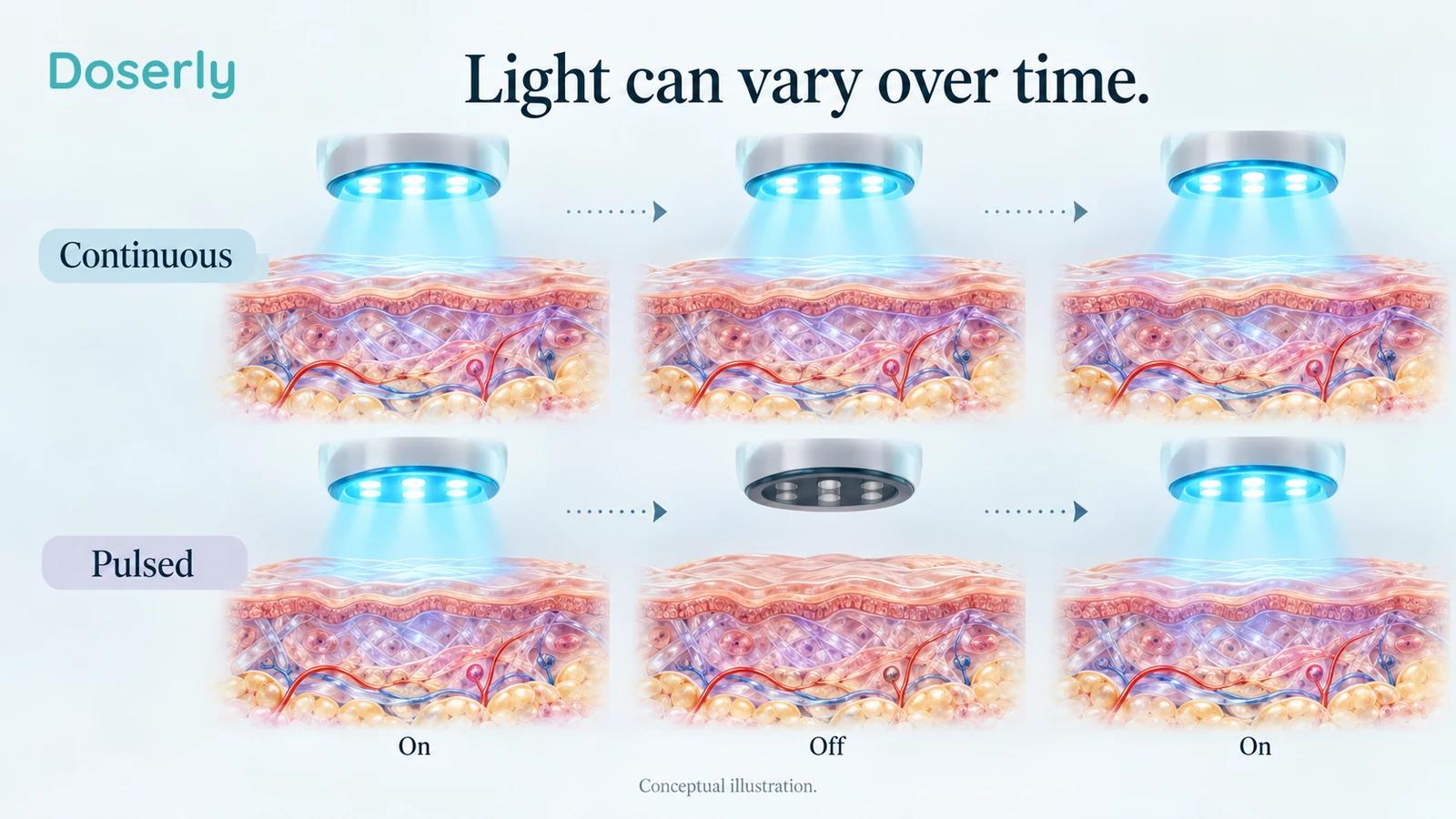
A pulsed source alternates between on and off periods. Its peak output during an on period can differ substantially from its average output over the full session. The duty cycle describes how much of that time the source is on.
For an ideal rectangular pulse pattern, a 50% duty cycle means the source is on half the time. If the on-state irradiance were 40 mW/cm² and the off-state output were zero, the average would be 20 mW/cm². These are illustrative numbers, not a clinical setting. More complicated waveforms require more complete characterization.
The distinction affects any energy calculation. Multiplying peak irradiance by total session time as though the source were continuously on overstates exposure in that example. Conversely, assuming every advertised number is a peak can also be wrong. The specification should say what was measured.
Pulse frequency alone does not tell you the energy delivered. A statement that a frequency resembles a brain rhythm is a hypothesis about biological relevance, not evidence that the device improves memory or mood. Wavelength, average output, pulse width, target and outcome remain part of the question.
A 2026 randomized study involving 31 people with major depressive disorder compared acute transcranial exposures, including continuous and pulsed conditions, with sham. Brain blood-oxygenation signal power changed differently across conditions, but single administrations did not significantly improve depression severity versus sham. The study is interesting for physiological dose research, not a basis for choosing a home pulse setting. Device-related commercial and intellectual-property relationships were disclosed.
Nonthermal does not mean temperature can be ignored
PBM is usually discussed as an approach intended to influence biological responses without deliberately destroying or ablating tissue. That is different from a laser treatment designed to heat a vessel, remove hair or resurface skin. The wavelength alone cannot tell you which kind of intervention is being delivered.
Temperature nevertheless remains relevant. A light source can warm skin even when heating is not its intended therapeutic mechanism. Device contact, exposure duration, ventilation and energy distribution influence comfort. An experiment that does not measure temperature carefully may have difficulty separating photochemical and thermal contributions.
Controlled red LED skin-safety trials observed dose-limiting adverse skin responses at high exposures, including blistering or persistent redness. Their forearm dose-escalation design does not establish a universal home-use limit. It does establish that “red” and “non-ionizing” are not synonyms for unlimited safety.
Warmth is therefore not proof that a useful dose has reached a deep target. Lack of warmth is not proof that exposure is appropriate for the eyes. Neither sensation substitutes for device-specific safety information, particularly when an application was never tested at the body site being considered.
Older popular explanations can blur these categories. In Huberman Lab's earlier light-and-health episode, the published transcript describes some skin effects using language about mild burning and removal of surface layers. That should not be carried over as an explanation of routine nonablative PBM. The same transcript discusses eye exposure in terms of comfort; comfort alone should not be used to validate a consumer panel for ocular treatment. The page notes that its transcript may contain errors, and the actual clinical protocols remain the appropriate reference.
What the podcast glucose claim actually measures
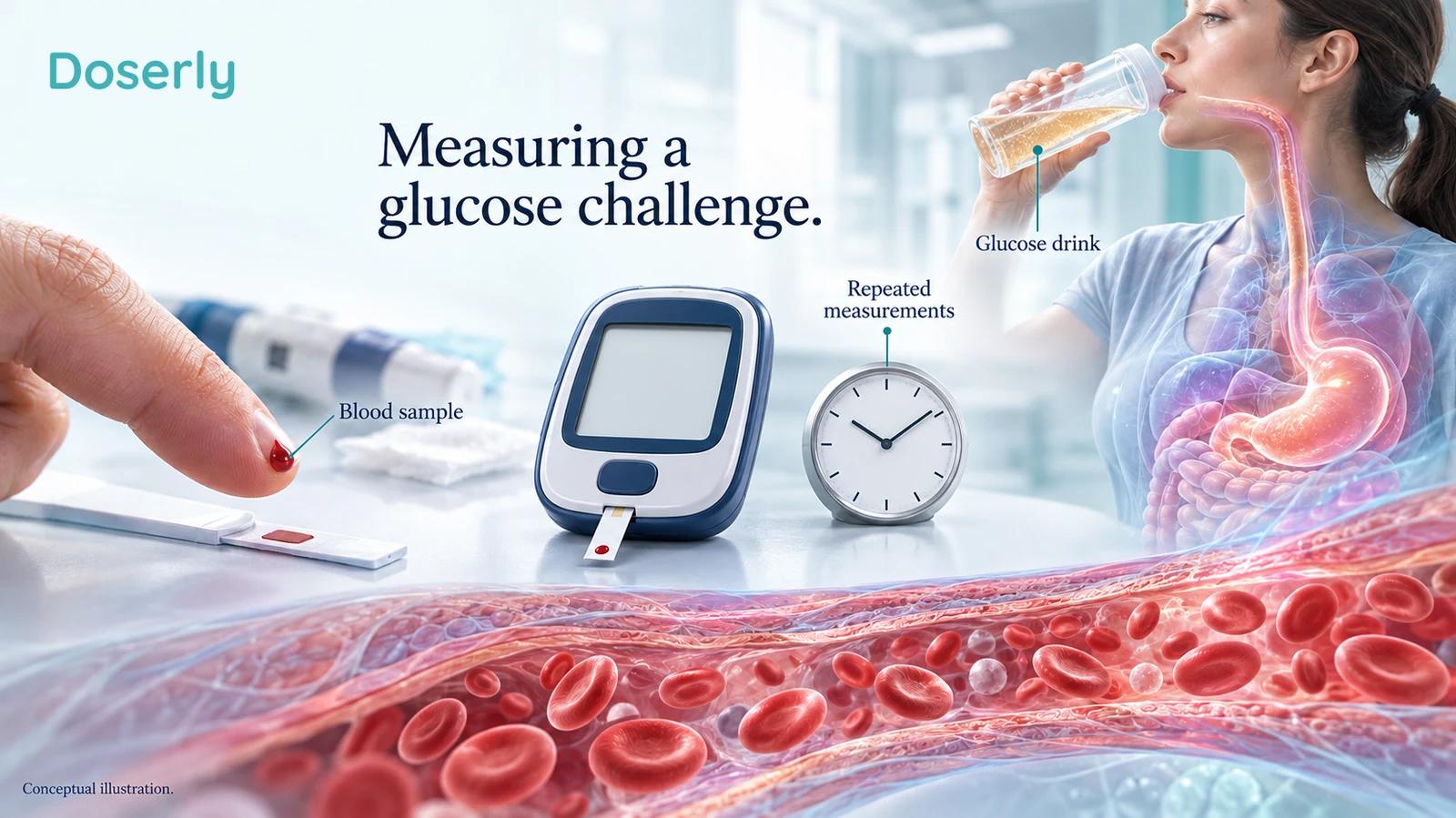
In the December 2025 Huberman Lab conversation with Glen Jeffery, the discussion moves from light transmission to blood glucose and possible systemic effects. The episode's official page also links its YouTube presentation. The episode includes a red-light-device sponsor, which is relevant commercial context rather than a reason to dismiss its research references.
The underlying 2024 Powner and Jeffery experiment recruited 30 healthy people, with 15 assigned to red light and 15 to a no-light comparison. After a glucose drink, the rise above baseline integrated over two hours was 27.7% lower with the tested intervention. The reduction in total glucose exposure was 7.3%. A separate comparison within the red-light group found a 7.5% lower peak than during those participants' earlier test without the intervention. Those percentages describe different calculations and comparisons.
Calling that a 27.7% reduction in blood sugar without the denominator obscures the result. It was an acute glucose challenge in people without known metabolic disease, not evidence of a sustained 27.7% reduction in everyday glucose, HbA1c or diabetes complications. The trial is a reason for further investigation; it does not establish a replacement for diabetes care or a transferable home protocol.
The distinction is useful beyond this one study. A change in a physiological challenge test can reveal an effect while leaving durability, clinical benefit and generalizability unresolved. The precise endpoint should travel with the impressive percentage whenever it is repeated.
Vision research needs equally precise language
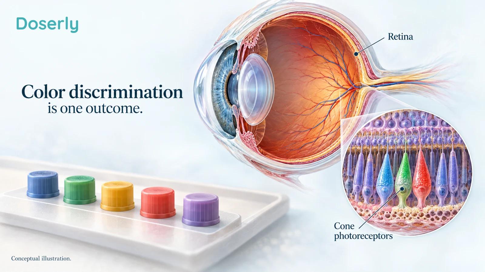
“Improves eyesight” can mean reading smaller letters, distinguishing colors, detecting contrast, preventing disease or navigating daily life more easily. These are different outcomes. An experiment can improve one without demonstrating the others.
The 2021 study of brief 670 nm exposures measured color-contrast thresholds in healthy adults using small, partly overlapping cohorts. Morning exposure was associated with improved thresholds, with follow-up extending to a week in a subgroup; the investigators also included a no-light control cohort. That is more specific than reversing all age-related vision loss. It does not establish a safe do-it-yourself eye treatment with an arbitrary red lamp.
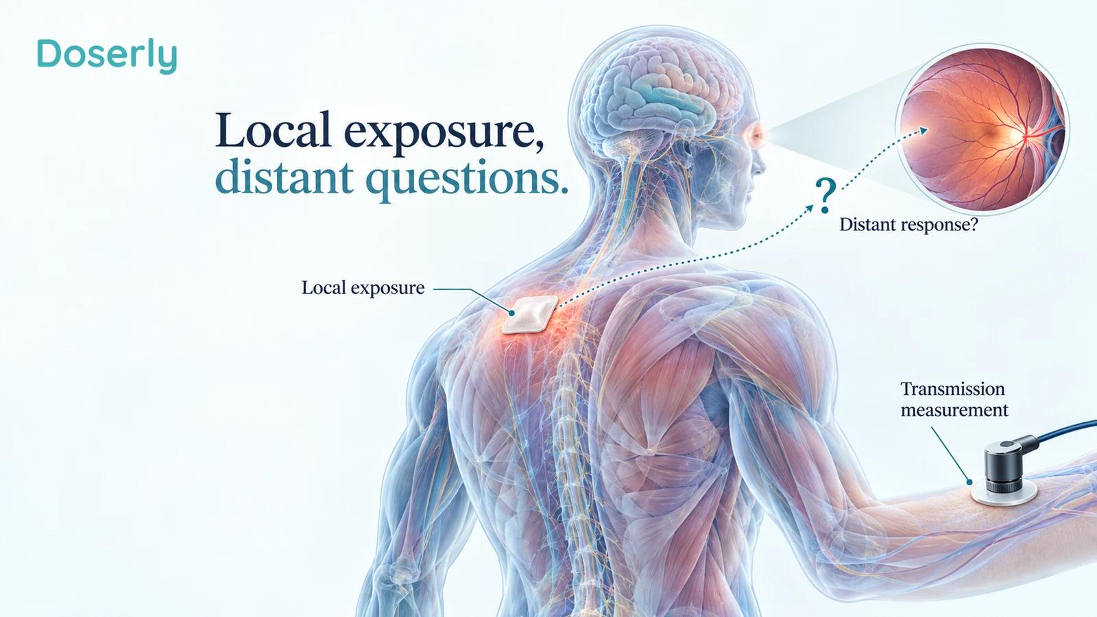
A 2025 study of light transmission and visual function included several small human experiments. Near-infrared light was detected after transmission through the thorax, and a separate experiment examined color-contrast thresholds after exposure to the back. A head-shielded subgroup contained only five people. Its blue-yellow threshold result was statistically significant, while its red-green result was not. Small, differently composed groups and short follow-up limit broad conclusions about systemic effects.
Detecting transmitted light and observing a remote outcome are both interesting. They do not prove that light directly illuminated every organ at an effective dose, or identify the signaling route responsible for the outcome. A remote effect, if replicated, might involve processes other than direct delivery to the measured organ.
In September 2026, Robinson and colleagues reported a randomized, single-blind study of 55 healthy adults comparing desk LED lighting, LED lighting supplemented with 850 nm and a control environment over one week. Color-arrangement error scores worsened in the LED group and improved in the supplemented group. Assessors were not blinded. The endpoint was color discrimination, not retinal injury, visual acuity, systemic mitochondrial health or lifespan.
This newer result deserves replication with careful exposure measurement and longer follow-up. It should neither be ignored because it is surprising nor expanded into a claim that ordinary lighting causes broad disease. Its relevance remains tied to the measured task and tested conditions.
Indoor lighting, sunlight and therapy are different exposures
The Jeffery podcast also makes broad claims about ordinary LED lighting and mitochondrial health. Those claims extend beyond the clinical outcomes established by the small studies discussed above. The research questions are legitimate; the certainty should remain proportional to the design and endpoint.
The International Commission on Illumination's position on blue-light hazard distinguishes retinal photochemical injury from sleep and circadian effects. It does not consider ordinary white general-lighting sources a blue-light hazard under reasonably foreseeable use. That statement addresses a defined safety question; it does not settle every emerging question about long-term lighting environments or color-discrimination performance.
Likewise, sunlight is not a calibrated PBM device. It includes a broad spectrum, changes with weather and time, and reaches different body surfaces under different conditions. Observational associations involving time outdoors cannot isolate red wavelengths from exercise, environment, social behavior or other exposures. They do not supply a reason to add ultraviolet exposure to a PBM routine.
A 2025 randomized crossover study in women investigated acute resting energy expenditure after a combined red and near-infrared intervention. The study included 16 women with obesity and 16 normal-weight sedentary women. An acute metabolic measurement is not evidence of sustained weight loss or longer life, and it does not determine which wavelength contributed to the response.
More recent exploratory work also needs that boundary. A 2026 uncontrolled pilot in people with type 2 diabetes reported some glycemic changes after a multiwavelength intervention but explicitly cautioned that its small sample and lack of a control group did not establish efficacy or causality. A newer publication date does not remove those design limits.
What public experiences can and cannot add
Community discussions are useful for understanding how people experience devices and where instructions become confusing. They are much less reliable for determining whether a particular wavelength caused a health improvement.
In a public December 2025 red-light-therapy discussion, an anonymous bed owner described feeling better and finding sessions relaxing, while openly listing many simultaneous changes, including exercise, weight-loss treatment and skin care. The author could not confidently separate the bed's contribution. That is a candid account of an attribution problem, not a controlled efficacy result. The post's additional drug and supplement practices are not recommendations.
Another public discussion of the glucose study included a participant asserting that 660 nm should work because it is equivalent to 670 nm. This is a community claim, not a demonstrated head-to-head clinical result. Nearby wavelengths may have similar properties in some contexts, but translating a study requires the rest of the exposure and a relevant outcome comparison too.
The useful response to these stories is neither automatic belief nor automatic dismissal. A person may sincerely notice better comfort or appearance. Without a stable baseline and comparison, however, it is difficult to know what produced the change. Keeping track of other treatments, activity, photographs and timing makes an account easier to interpret, even though it does not turn an individual experience into a randomized trial.
Why stronger and more frequent can still fail
Dose-response relationships are not guaranteed to be linear. A review of biphasic PBM responses describes varied experimental evidence in which increasing exposure does not continually increase the response. Much of that evidence comes from laboratory systems. It cannot identify one universal optimal window for every human tissue.
Clinical dose comparisons also sometimes find no useful effect. In a 2024 placebo-controlled crossover study of 18 runners, three tested total-energy conditions did not improve 5 km performance or perceived exertion compared with placebo. This does not answer every sports question, but it directly challenges the assumption that a larger energy total must produce a better result.
When an outcome remains uncertain, increasing frequency is not a substitute for evidence. Nor is combining more wavelengths, adding heat or changing distance until the session feels stronger. Those changes alter the intervention and may make it less comparable with the research being cited.
The most informative future studies will compare clearly characterized exposures, use credible controls, report adverse effects and follow outcomes beyond the treatment period. For readers now, the best available decision is to match the complete tested intervention to a specific goal and remain honest about the size of the remaining gap.
A practical comparison that stays close to the evidence
When reading a paper or device specification, begin with the treatment goal and then work outward. The wavelength should be one part of a description that includes the target tissue, light source, measured exposure, treatment area and schedule. The outcome should be something the study actually measured.
Specification or claim
“660 nm for skin”
- A useful follow-up question
- Which skin condition, device and clinical outcome were studied?
Specification or claim
“850 nm reaches deeper”
- A useful follow-up question
- Deeper than what, measured how, and with what remaining exposure?
Specification or claim
“Multiple wavelengths work together”
- A useful follow-up question
- Was the combination compared with its individual components?
Specification or claim
“Laser-grade treatment”
- A useful follow-up question
- What does that mean for this device's output, coverage and intended use?
Specification or claim
“Mitochondrial activation”
- A useful follow-up question
- Was that measured directly, inferred from a signal, or used as an explanation?
Specification or claim
“Clinically studied wavelength”
- A useful follow-up question
- Was the actual device and complete treatment method studied?
These are not obstacles to using the evidence. They are ways to make the evidence more relevant. A manufacturer that can explain what was measured and what remains uncertain is giving you more useful information than one that relies on a fixed-depth diagram and a list of impressive numbers.
For clinicians and researchers, complete reporting serves the same purpose. If wavelength, treatment geometry or output measurement is missing, a seemingly similar intervention may not be reproducible. A 2025 reporting proposal distinguishes output at the device aperture from irradiance at the target surface and emphasizes beam profile and delivery details. Updated laser-therapy reporting proposal.
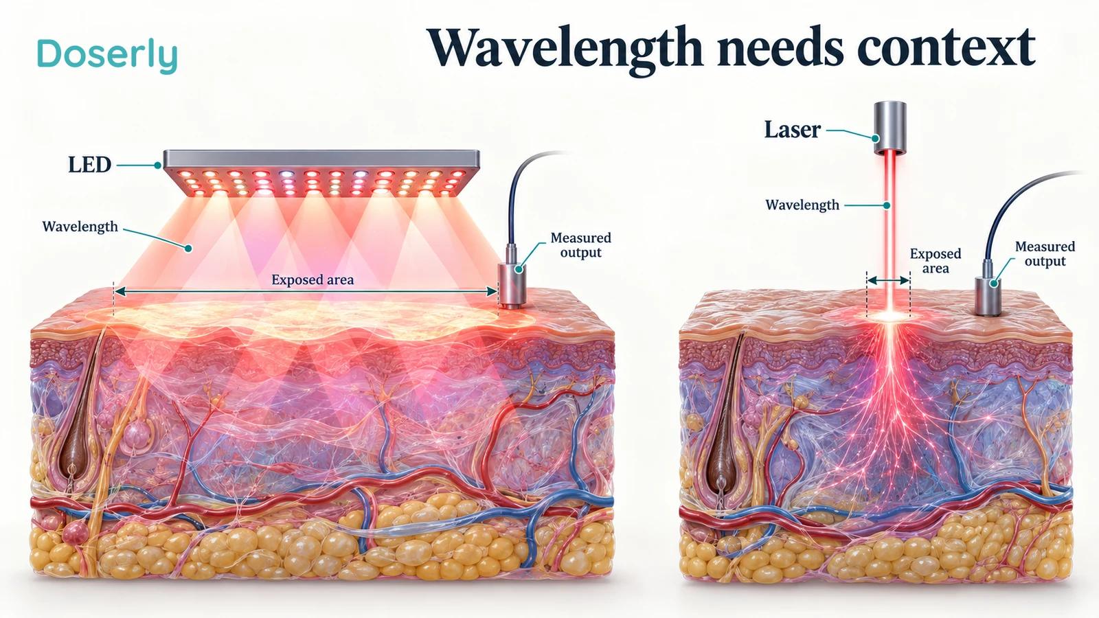
Red and near-infrared light can behave differently in tissue, and those differences are scientifically useful. The strongest conclusions remain tied to a particular question: which tissue, which exposure, which person and which outcome? A wavelength can help describe the treatment. It cannot, by itself, tell you what that treatment will accomplish.