
Peptides
October 16, 2026
Humanin and insulin sensitivity: what the metabolic studies show
On this page
Humanin has a credible metabolic research story, but it is easy to tell that story incorrectly. Experiments have examined insulin action in rats, pancreatic cell survival in laboratory models and naturally occurring Humanin concentrations in people with different glucose profiles. Those findings do not add up to a demonstrated Humanin treatment for insulin resistance in humans.
The distinction is practical. A lower peptide concentration in a diabetes cohort does not identify a deficiency that should be replaced. A glucose change in an animal does not establish the safety or benefit of an injected preparation in a person. Understanding the actual endpoints makes the research more informative without turning it into a regimen.
The Humanin guide provides the wider background. Here, the central questions are what insulin sensitivity means, which experiments measured it, and why pancreatic protection and blood-marker associations belong in separate parts of the explanation.
Insulin sensitivity is a response, not simply a glucose number
Insulin helps coordinate glucose handling across tissues. Insulin resistance means that tissues such as muscle, liver and fat respond less effectively to it. Blood glucose can reflect both that response and the pancreas’s ability to supply insulin. The National Institute of Diabetes and Digestive and Kidney Diseases distinguishes insulin resistance from the glucose-based tests used to identify prediabetes. NIDDK explanation.
A single glucose concentration therefore cannot identify every mechanism behind it. Two people with similar readings may differ in insulin secretion, tissue response, medication exposure or the circumstances of the sample. A study claiming improved insulin sensitivity needs an assessment designed for that question, rather than assuming that any reduction in glucose proves the same mechanism.
This also separates sensitivity from pancreatic beta-cell survival. Beta cells produce insulin. Preserving those cells could be valuable in a suitable disease context, but it is a different claim from making muscle or liver respond more strongly to insulin already present. Humanin research has investigated both ideas; keeping them separate prevents one promising experiment from answering two questions at once.
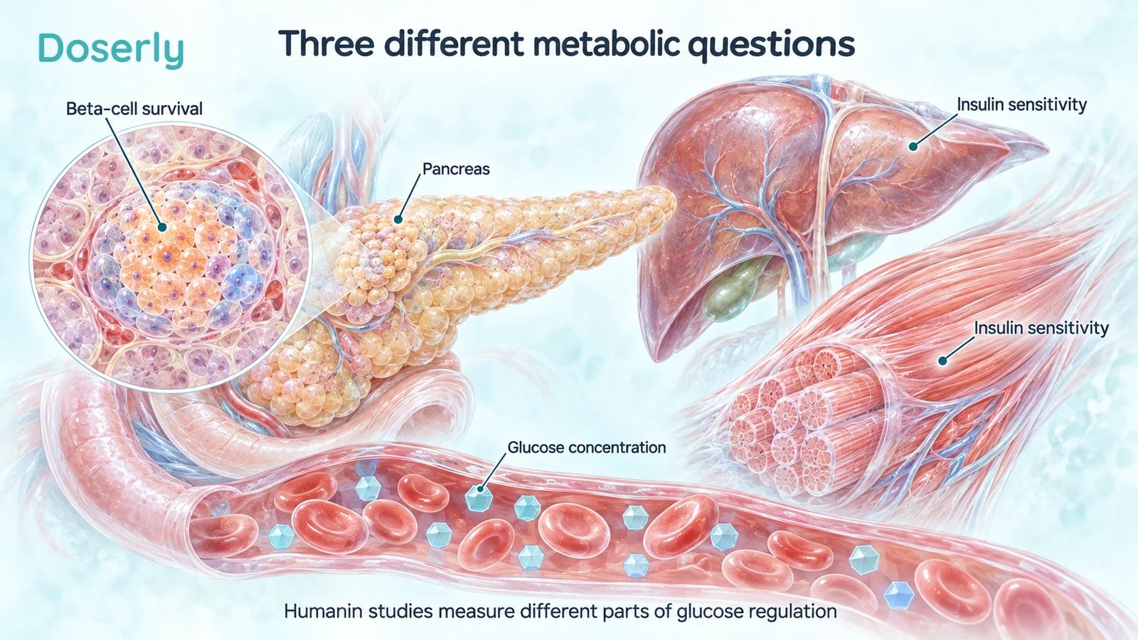
Beta-cell survival, insulin sensitivity and glucose concentration are related but distinct endpoints. The organs illustrate the questions being studied, not proven Humanin benefits in patients.
The insulin-action experiments were in rodents
In a 2009 PLOS ONE paper, Muzumdar and colleagues used controlled metabolic experiments in rats. Central Humanin infusion and peripheral administration of a potent analogue, HNGF6A, improved insulin action under the study conditions. The investigators examined hypothalamic STAT3 signaling and found that inhibiting it blocked important metabolic effects. A separate experiment reported lower glucose after an analogue treatment in Zucker diabetic fatty rats. Original insulin-action study.
That is more mechanistically informative than a simple before-and-after glucose observation. The study manipulated the candidate pathway and tested whether the response depended on it. Nevertheless, the result remains specific to the animal models, molecules and delivery methods used. It does not establish that an ordinary peripheral Humanin injection reproduces central infusion or HNGF6A exposure in a human.
The distinction between native Humanin and HNGF6A is especially important. An analogue is a deliberately altered molecule. Its study is not a clinical validation of every product bearing the Humanin name. A claim about one sequence needs evidence about that sequence, including the formulation and route through which it was administered.
The word “central” also has a concrete meaning here: the investigators were examining brain-mediated regulation of peripheral metabolism. It should not be translated into a general claim that Humanin repairs the brain or improves every metabolic organ directly. The experiment was designed to identify signaling relationships, not to demonstrate whole-body rejuvenation.
Why a clamp is more informative than an isolated sample
The 2009 work used clamp methods to examine insulin action. In a metabolic clamp, researchers control the experimental conditions and monitor how glucose must be supplied to maintain a specified blood-glucose state during insulin exposure. With additional measurements, the design can help distinguish changes in glucose production from changes in glucose uptake. That is the purpose of using a controlled physiological experiment rather than relying only on a resting concentration. Clamp study methods.
The general lesson is about matching a measurement to a claim. If a treatment is said to improve insulin action, measuring the response to insulin is more direct than observing an associated peptide. If it is said to prevent diabetes complications, even a convincing short-term sensitivity result would leave a much longer clinical question open.
A mechanistic result is therefore valuable without being the final clinical endpoint. It can guide candidate selection, identify a pathway to investigate and help design a human study. It cannot by itself tell someone whether treatment will improve their daily health, how long an effect lasts, or whether another organ experiences an unwanted effect.
Pancreatic-cell protection addresses a different problem
Hoang and colleagues studied Humanin in mouse-derived beta cells and nonobese diabetic, or NOD, mice. They reported reduced cell death under specified laboratory stresses and findings consistent with delayed or improved diabetes outcomes in the mouse model. This research focused on beta-cell survival and a model relevant to autoimmune diabetes. It was not a trial of Humanin in people with type 2 diabetes. Beta-cell and NOD-mouse study.
The model distinction matters because “diabetes” is not one interchangeable experimental condition. A model of immune-associated beta-cell loss does not reproduce every aspect of common type 2 diabetes. Likewise, protecting cultured beta cells from a selected challenge does not establish that a person’s own insulin production will recover.
There is no contradiction in a peptide being investigated for more than one mechanism. The problem arises when the mechanisms are merged into a stronger clinical claim than any experiment tested. “Investigated for beta-cell protection” and “improved insulin action in a rat clamp” are two specific statements. Neither should be shortened to “treats diabetes.”
A future clinical program would need to decide which problem it is trying to solve. Preserving insulin secretion early in one disease, improving insulin sensitivity in another and reducing long-term complications would require different populations and outcomes. The broad biological interest does not remove that need for a precise clinical purpose.
What the human blood studies add
A 2016 observational study analyzed 81 participants, including people with impaired fasting glucose and controls. Its results table placed lower plasma Humanin in the impaired-fasting-glucose group. The abstract’s numerical group labels are inconsistent with that table, so the result should be read from the full results rather than copied mechanically. No Humanin treatment was assigned. Primary study, Table 2.
A 2019 study compared 225 people across control, prediabetes and type 2 diabetes groups and found lower serum Humanin in the dysglycemic groups. The authors noted that samples were collected randomly, preventing calculation of fasting-based insulin-resistance indices such as HOMA-IR. The control group was also younger, and participants were overweight or obese. These are material limits to interpreting the association. Diabetes cohort and limitations.
These studies connect Humanin measurements with human metabolic conditions. They do not establish that low Humanin caused the condition, that increasing it reverses the condition, or that the measured concentration provides a personal dosing target. Those are intervention questions that observational group comparisons cannot settle.
An association can still be useful. It may help generate a hypothesis about disease biology or identify a candidate biomarker for further validation. The value comes from stating exactly what the comparison contributes, rather than treating observational evidence as disappointing because it is not already a treatment trial.
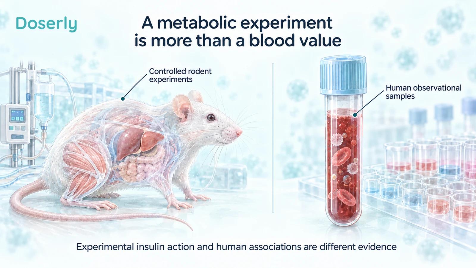
Rodent intervention experiments and human sample comparisons supply different evidence. This conceptual illustration contains no dosing instructions or implied human treatment result.
The human findings are not uniform
A larger study comparing mitokines across healthy aging, type 2 diabetes and Alzheimer’s disease reported that Humanin was lower in Alzheimer’s disease but not in the type 2 diabetes group relative to the relevant comparison. This differs from a simple story in which Humanin is always low whenever glucose regulation is impaired. Disease-specific mitokine study.
Differences between studies can reflect populations, disease stages, assays, specimen types or other design choices. The existing comparisons do not allow every discrepancy to be assigned to one cause. It would be equally unwarranted to dismiss all associations as laboratory error or to assume that each study reveals a universal biological rule.
For a reader, this means resisting the temptation to choose whichever study gives the cleanest narrative. A biomarker intended for clinical use has to perform across the settings in which it will be used. If its relationship to disease varies, researchers need to understand that variation before defining a threshold or recommending action on an individual result.
The same caution applies to a claim that a peptide is “depleted.” That word implies a known desirable reserve and an established deficit. A lower group average, even when reproducible, does not establish either. Describing the actual measurement is more accurate than importing the logic of a validated deficiency condition.
Exercise observations do not identify a replacement therapy
A resistance-training study found increased Humanin protein in muscle from men with impaired glucose metabolism without a corresponding serum increase. It did not establish a consistent relationship between individual Humanin changes and improvements in metabolic or fitness measures. The intervention was exercise, not administered Humanin. Training study.
This is a useful boundary for mechanistic explanations of exercise. A molecule can change during an intervention without being the sole cause of the intervention’s effects. To establish mediation, researchers would need evidence connecting the molecule’s change to the outcome and distinguishing it from other adaptations.
The observation also cannot be reversed into the claim that injecting the molecule reproduces training. Exercise changes many processes together, with timing and tissue patterns that a preparation may not reproduce. A candidate pathway can be scientifically interesting while remaining insufficient as a substitute for the activity that first revealed it.
What would advance the treatment question?
The next meaningful step would be evidence for a defined candidate in an appropriate human population. Early studies would need to establish exposure and tolerability. A controlled efficacy study would then need outcomes suited to its claim: a validated assessment of insulin action, sustained glucose control, or a clearly specified clinical benefit.
The comparator and background treatment would matter. People receiving diabetes care may already use interventions that change glucose levels. A study would need to account for those treatments, changes in body weight, diet and activity, and the possibility of adverse effects. An improvement should be attributable to the candidate rather than to simultaneous changes or selective reporting.
Longer-term claims would require longer-term evidence. A short physiological experiment cannot establish prevention of kidney, eye or cardiovascular complications. Nor would a favorable biomarker change resolve the benefit-harm balance of chronic exposure to a cell-survival signal. Those questions need to be planned and measured, not inferred from the attractiveness of the mechanism.
Use established assessment for a real glucose concern
For current clinical decisions, NIDDK describes fasting plasma glucose, A1C and oral glucose-tolerance testing as established ways to assess diabetes or prediabetes in the appropriate context. The tests answer related but different questions, and clinicians interpret them alongside history and other results. Humanin measurement is not presented as a replacement for that assessment. NIDDK testing guidance.
A useful conversation therefore starts with the actual concern: an abnormal result, a trend, symptoms, family history or a medication change. That gives a clinician something specific to evaluate. An experimental Humanin result cannot tell you on its own whether established treatment should change.
Humanin’s metabolic research supports further investigation of a biologically interesting pathway. Its strongest intervention findings here come from controlled animal experiments, while the human evidence mainly concerns associations and responses to other interventions. Preserving that distinction is what allows the research to inform curiosity without becoming unsupported treatment advice.
Sources
- Muzumdar et al., 2009: central and peripheral insulin action.
- Hoang et al., 2010: beta-cell protection and NOD mice.
- Voigt and Jelinek, 2016: impaired fasting glucose.
- Ramanjaneya et al., 2019: diabetes cohort.
- Conte et al., 2021: disease-specific mitokines.
- Gidlund et al., 2016: resistance training.
- NIDDK: insulin resistance and prediabetes.
- NIDDK: diabetes and prediabetes tests.