
Peptides
October 16, 2026
Humanin and exercise: what changes in muscle and blood mean
On this page
Exercise makes muscle do more than contract. It changes energy demand and activates signals involved in the body's response to that demand. Humanin, a small mitochondrial-derived peptide, is one of the signals researchers have measured around exercise. The practical question is whether it helps explain training adaptations—and whether any of this supports taking humanin to improve fitness.
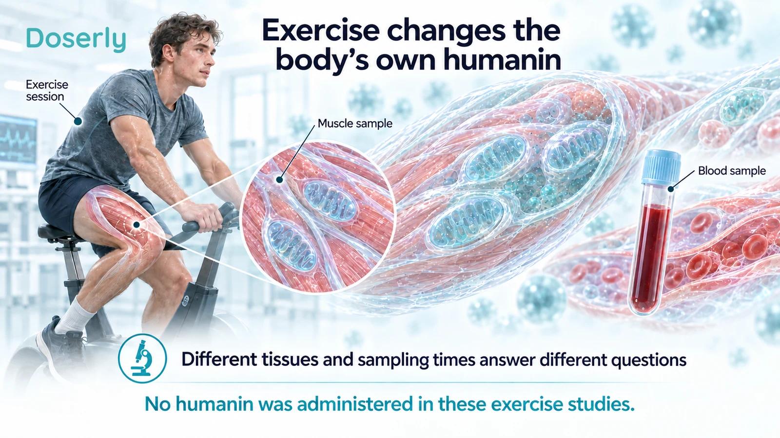
The human studies described here show that exercise can change the body's own humanin in muscle or blood. They did not give participants humanin. Their results also depend on whether researchers examined one session or weeks of training, which exercise was performed and which tissue was sampled. Those details explain why apparently different findings can all be reasonable. Acute exercise study, training study
There is no exercise-equivalent humanin regimen established by these studies. But they offer a useful view of how researchers investigate adaptation, and why a temporary rise in a peptide is not the same as a demonstrated improvement in strength or endurance.
Why look for humanin in muscle?
Skeletal muscle contains mitochondria that help meet its energy needs. Humanin has been investigated in cellular stress and metabolic research, making it a plausible candidate for a signal that changes when muscle works harder. Measuring it during exercise asks whether it responds under real physiological conditions, rather than only when a laboratory researcher exposes cells to a substance.
Researchers can collect a muscle biopsy, a small tissue sample, and a blood sample from the same person. The biopsy provides information about the sampled muscle. Blood provides information about what is circulating at that moment. These are connected compartments, but neither is a complete substitute for the other. Woodhead and colleagues
The distinction between endogenous and administered humanin is equally important. Endogenous means made within the body. Measuring it after cycling does not test the consequences of injecting additional peptide, and it does not establish that the measured change caused the benefits of cycling. Our humanin guide explains the native peptide and the modified analogues used in other experiments.
A 12-week resistance-training study
A 2016 report examined men with impaired glucose regulation, a metabolic state relevant to diabetes risk. The parent randomized exercise study included resistance training, Nordic walking and a control group. A subgroup of 55 men donated muscle samples; usable numbers differed by assay. For the muscle humanin protein analysis, the figure reported 15 resistance-training, 14 Nordic-walking and 13 control participants.
After 12 weeks, muscle humanin increased in the resistance-training group, by approximately 1.35-fold in the reported analysis. The other groups did not show the same significant change. Serum humanin did not change significantly over time or between groups. Gidlund and colleagues
This is a particularly useful result for understanding the topic. It supports a muscle response to training in the population studied, while showing that a blood test may not mirror it. Describing the finding simply as “exercise raises humanin” loses the tissue-specific result.
The investigators also reported differences in baseline muscle values between groups and accounted for them statistically. Changes in muscle humanin did not consistently track improvements across the metabolic and fitness measurements. The study therefore did not establish that humanin was the mechanism responsible for improved glucose handling or fitness.
For a reader with prediabetes, the participants are more relevant than healthy laboratory animals. Even so, the intervention was exercise. The study cannot tell us whether administering a peptide produces the same adaptations, whether a particular blood level should be targeted or whether resistance training is universally better than walking.
One bout of high-intensity cycling
A 2020 study investigated a shorter time scale in 10 healthy, untrained young men, whose average age was about 25. They completed ten one-minute cycling intervals at a workload based on their peak oxygen uptake. Researchers collected muscle and plasma before exercise, immediately afterward and after four hours. They repeated testing after six interval-training sessions over two weeks.
Humanin increased in muscle and plasma after the acute exercise session. Following the short training period, resting plasma values showed a downward trend that did not meet the conventional statistical threshold: P=0.063. The acute response was not eliminated by training. Woodhead and colleagues
The result does not mean training “used up” humanin or made the participants deficient. A resting concentration and the response to a new bout of exercise answer different questions. The downward trend also needs to remain a trend, rather than being rewritten as an established decrease.
The participant group was small and restricted to young men. That limits what can be inferred about older adults, women, people with chronic disease or very different training histories. The published exercise workload describes how the study was performed; it is not a prescription for someone trying to maximize humanin.
Endurance and resistance exercise in the same experiment
A 2021 study randomized 30 participants, ten each, to endurance exercise, resistance exercise or a control condition. The endurance session involved 45 minutes of cycling at 70% of estimated maximal oxygen uptake. The resistance session used leg press and knee extension. Blood and muscle were sampled before and at 30 minutes and three hours afterward.
Circulating humanin increased after endurance exercise but not significantly after the resistance session. The related peptide MOTS-c showed a trend after endurance exercise. At rest, the investigators did not find that these peptide measurements tracked the fitness outcomes they assessed. Primary report
That doesn't overturn the 12-week resistance-training result. One study measured an immediate blood response to a single session; the other found a muscle-protein change after repeated training in men with impaired glucose regulation. The time scale, population and sample differ. A statement about one cannot simply be substituted for the other.
Sample availability also reduced the number of analyses for some measures in the acute study. The number randomized should therefore not be mistaken for the number contributing to every laboratory comparison. This is especially important in small mechanistic studies, where missing a few samples can materially affect precision.
Why blood and muscle need not move together
A muscle concentration reflects the amount detected in the sampled tissue under the assay conditions. A circulating concentration reflects the balance of release, distribution and removal across the body. It may have contributions from multiple tissues. These are reasons to expect that the two measurements can behave differently, not proof that muscle is the sole source of a blood change.
The 2020 study included a separate experiment in isolated mouse muscle, where contraction increased measured humanin. That supports investigating muscle as a contributor. It does not by itself quantify how much of the circulating change in exercising people came from their muscle. Woodhead and colleagues
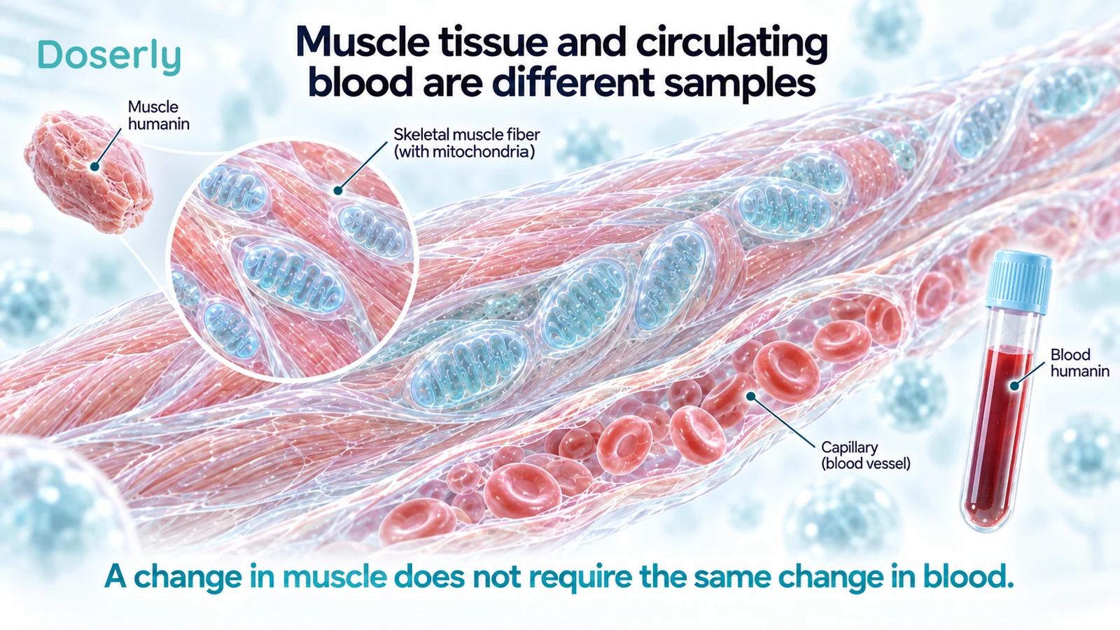
Timing adds another layer. A sample taken immediately after exercise might capture a transient response that a sample taken the next morning misses. Conversely, a resting sample after weeks of training might reveal an adaptation that is different from the acute response. Without timing information, comparing two numbers can be misleading even if the laboratory method is identical.
This is why a higher post-exercise reading should not be treated as a score of workout quality. The studies did not establish a target rise that predicts greater strength, better glucose control or faster recovery.
Does the diabetes literature make humanin a treatment target?
A separate cross-sectional study measured mitochondrial-derived peptides in 225 people with normal glucose regulation, prediabetes or type 2 diabetes. It found lower serum humanin in the dysglycemia groups and relationships with glucose-related measurements. The participants were not treated with humanin. Ramanjaneya and colleagues
The study gives metabolic context for exercise research, but it doesn't identify a correctable humanin deficiency. Lower levels could reflect disease, contribute to it or accompany another process. The authors noted that controls were younger and that the sampling did not allow calculation of certain fasting indices of insulin resistance.
Combining that association with the training study generates a hypothesis: perhaps humanin participates in some of the metabolic response to exercise. Testing that hypothesis would require demonstrating the role of humanin itself, rather than assuming that two associated findings complete the causal explanation.
For clinical decisions, glucose regulation and physical function remain the meaningful questions. The reviewed studies do not show that choosing exercise or treatment according to a humanin test improves those outcomes.
What the 2026 muscle-cell paper adds
A newer paper examined primary human muscle cells grown in culture, not people exercising or receiving injections. Researchers exposed the cells to dexamethasone, a glucocorticoid used to model muscle atrophy, and separately tested HNG/S14G-humanin or MOTS-c. HNG is an analogue, not native humanin.
HNG preserved the measured myotube area under the experimental conditions, but did not reproduce every effect observed with MOTS-c. For example, the report distinguished effects on the fusion index and markers involved in protein breakdown. Elhusseiny and colleagues, 2026
Myotubes are developing muscle-cell structures in culture. Their area and molecular responses are useful experimental outcomes, but they are not measurements of leg strength, exercise recovery or preserved independence in patients. The word human in “human muscle cells” identifies the cells' origin; it does not make the experiment a human treatment trial.
The study also concerns drug-induced atrophy, not the same physiological setting as an ordinary workout. It helps researchers investigate mechanisms worth pursuing while leaving the whole-person treatment question open.
Why an injection is not a copy of an exercise response
An injected peptide has a dose, formulation, route and exposure pattern that need independent characterization. The body's response during exercise is changing alongside circulation, fuel use and many other signals. Reproducing one measured concentration would not demonstrate that the rest of that response had been reproduced.
A pharmacokinetic study in male rodents found differences in the behavior of HNG and HNGF6A, another modified analogue, and differences between rats and mice. Pharmacokinetics describes how a substance is distributed and cleared over time. These results underline why a rodent half-life or analogue result should not be converted into a human exercise protocol. Chin and colleagues
The exercise studies discussed above provide no validated dose for that purpose. They also do not establish the safety of repeated humanin administration around training or of combining it with other peptides.
What should you take from the exercise findings?
Humanin appears to be exercise-responsive under several studied conditions. The most useful details are the distinction between muscle and blood, the difference between a single bout and repeated training, and the fact that participants were not given humanin. Those details explain the evidence more faithfully than a general claim that it is an exercise mimetic.
If a training or treatment proposal cites these papers, ask which outcome it relies on. A muscle-protein change after 12 weeks, a transient plasma rise and a cultured-cell response are different findings. None supplies a proven shortcut to strength or endurance.
For your own records, it is more useful to describe the actual training, symptoms, recovery and relevant clinical measurements than to assign meaning to an unvalidated peptide target. The research may eventually clarify humanin's contribution to adaptation. At present, it shows a physiological response worth studying, not an established replacement for exercise.
Sources
- Gidlund et al., resistance training and muscle humanin, 2016.
- Woodhead et al., high-intensity exercise, 2020.
- Acute endurance and resistance exercise study, 2021.
- Ramanjaneya et al., diabetes biomarker study, 2019.
- Elhusseiny et al., HNG and MOTS-c in cultured human myotubes, 2026.
- Chin et al., analogue pharmacokinetics in male rodents, 2013.