
Peptides
October 16, 2026
ARA-290 and corneal nerve imaging: what an eye scan can tell us
On this page
ARA-290, also called cibinetide, has been studied as a possible treatment for small-fiber neuropathy: damage to the thin nerves involved in sensation and automatic body functions. For someone living with burning feet, tingling or pain from light touch, an eye examination may seem an unexpected way to investigate treatment. Why photograph the cornea when the symptoms are somewhere else?
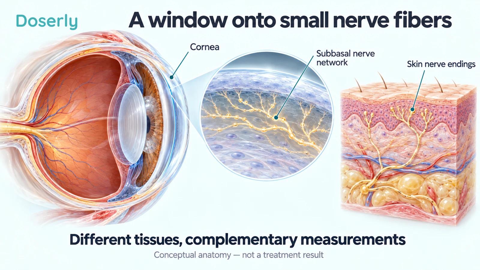
The cornea is the clear surface at the front of your eye. It contains a network of small sensory nerve fibers that researchers can examine in a living person using a specialized microscope. That accessibility makes it useful for studying nerve disease. It also creates an important distinction: a change in corneal nerves is a structural finding, while less pain or better walking is a clinical result. ARA-290 studies have measured both, with results that do not always move together.
This article explains what the pictures measure, why researchers use them, and what the human trials actually established. Our ARA-290 guide covers the compound's wider background and safety questions.
Why small nerves are difficult to study
Small nerve fibers extend into tissues where they detect temperature and potentially harmful stimuli. Damage can produce a mixture of reduced sensation and unpleasant sensations, rather than simply making an area numb. In sarcoidosis, an inflammatory disease, small-fiber symptoms can be a substantial problem even when the person's other disease measurements do not explain how unwell they feel.
Researchers therefore need several ways to examine the problem. Questionnaires record the person's experience. Sensory testing measures responses to controlled stimuli. Skin biopsies allow nerve fibers to be counted in a small tissue sample. Corneal confocal microscopy provides another view: detailed images of nerve fibers beneath the cornea's surface. The 2013 ARA-290 trial deliberately combined these approaches instead of treating any one of them as a complete description of neuropathy. Dahan and colleagues, methods
Each test has a location and a purpose. A skin biopsy samples a particular part of a leg or thigh; an eye image samples a particular region of the cornea. Neither is a photograph of every small nerve in the body. Agreement between different measurements can strengthen a finding, but disagreement needs explanation rather than being hidden.
What happens during corneal confocal microscopy?
“Confocal” describes an imaging method that isolates a thin optical section, allowing tiny structures to be seen with less interference from surrounding tissue. In the 2013 trial, researchers used topical eye anesthetic and positioned a specialized microscope at the cornea. They obtained images at successive depths, then analyzed the subbasal nerve layer near the surface. Each image covered a very small field, approximately 0.4 by 0.4 millimeters. Trial imaging methods
The result is not an ordinary photograph of the eye. It shows fine branching structures that an analyst or computer program can trace. Which images are chosen, how well the nerves are visible and how the tracing is performed all affect the final measurement. Researchers reduce bias by keeping image assessors unaware of treatment assignment and applying the same procedures across groups.
This also explains why a striking before-and-after picture cannot stand in for the whole trial. A published example may illustrate a particularly clear response. The treatment comparison needs the measurements from the analyzed participants, including people with little change, rather than the most impressive pair of pictures.
Repeated imaging can be useful because it does not require removing a new piece of tissue each time. That practical advantage helps researchers observe changes over relatively short periods. It does not remove the need for consistent sampling or establish that the sampled tissue represents every painful body region.
Fiber density, length and area are different measurements
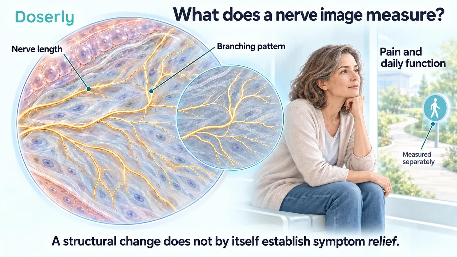
The phrase “more nerves” can conceal several different calculations. Nerve fiber density may count main nerve fibers within a specified image area. Nerve fiber length adds up the length of visible fibers. Nerve fiber area estimates how much of the image is occupied by the traced nerves. Branch density examines branching separately.
These measures are related, but an increase in one is not automatically an equal increase in another. Existing fibers may extend or branch without the number counted by a particular method changing much. Image-analysis choices also matter. The 2017 cibinetide trial selected corneal nerve fiber area as its main outcome; the diabetes trial used a nerve-density measure. Keeping those names attached to the results prevents a false comparison between unlike numbers. Cibinetide trial; diabetes trial methods
An actual correction illustrates why units deserve attention. A 2016 erratum to the 2013 study reported an arithmetic error in the corneal area algorithm: the original absolute values needed multiplication by 2.26. The erratum also specified an additional factor for expressing the corrected values per square millimeter. Applying a common scaling factor does not change the percentage change, but it does change the absolute area. Old numbers should not be copied into a new graphic without checking that correction. Published erratum
What did the larger ARA-290 trial find?
The 2017 phase 2b report described 64 people with sarcoidosis-associated small nerve fiber loss and neuropathic pain. Participants were assigned to placebo or one of three cibinetide amounts for 28 days. These were research exposures, not an established treatment schedule.
The main endpoint was change in corneal nerve fiber area at day 28. Only the 4 mg group showed a statistically significant improvement over placebo: the adjusted difference was about 697 square micrometers, with a 95% confidence interval of 159 to 1,236. The comparisons for 1 mg and 8 mg did not reach statistical significance. The results therefore do not establish a simple rule that more drug produces a larger or more reliable effect. Culver and colleagues, 2017
The investigators also examined skin. Total intraepidermal nerve fiber density did not show a treatment-group difference, while a marker associated with growing nerve fibers, GAP-43, increased in the 4 mg group. These are different observations: a growth-associated staining measurement is not identical to a count of all fibers crossing into the epidermis. Full primary report
This combination gives researchers a reason to investigate biological activity. It does not show that every affected nerve was restored, or that a patient had recovered normal sensation throughout the body.
Did the eye changes match better symptoms?
Pain improved in all groups in the 2017 trial, including placebo. In the subgroup with moderate-to-severe pain, the 4 mg comparison favored treatment numerically but was not statistically conclusive: the reported P value was 0.157. That should not be rewritten as proven pain relief simply because the corneal endpoint was positive. Primary trial abstract
There were correlations between corneal changes and some other measurements, including a growth-associated skin marker and six-minute walking distance. A correlation means that participants with a larger change in one measurement tended to have a larger change in another. It can support a biological hypothesis, but does not show that the corneal change caused the walking change. Both could be influenced by other factors, and small-group correlations can be unstable.
For a patient, the practical questions remain direct: did pain become less intrusive, did sleep improve, did walking become easier, and did those changes last? Imaging can complement those answers. It should not replace them.
Why the earlier studies still matter
The 2013 trial involved 38 people with sarcoidosis and documented small nerve fiber loss. After 28 days, the active-treatment group had a reported median corneal area increase of 14.5% from baseline. Its symptom findings depended on what was measured: a small-fiber symptom questionnaire and pain interference favored treatment, whereas pain intensity improved similarly in the two groups. Skin fiber density did not change significantly. 2013 primary report
That pattern is more informative than describing the trial as simply positive or negative. Pain interference concerns how pain affects activities; intensity concerns how strong it feels. A person might manage daily life better without reporting a large change in intensity. Conversely, improvement on one questionnaire does not establish every proposed benefit.
The diabetes trial adds another distinction. Across the full analyzed group, the corneal nerve-density increase was not significant. A later subgroup analysis focused on participants whose starting density was below a specified normal-reference threshold and found a signal there. The paper explicitly described that analysis as post hoc, meaning it was performed after the original study planning. It is useful for designing a better-targeted future study, but less secure than a successful prespecified comparison. Diabetes trial
Does an imaging improvement last?
An endpoint measured after four weeks tells us about that time point. It does not establish permanent repair. The 2017 investigators reported that corneal nerve fiber area moved back toward baseline after treatment stopped. The tissue finding therefore needs to be discussed with its follow-up, rather than presented as a lasting restoration. Full trial report
Different outcomes can also have different timelines. In the earlier sarcoidosis study, some questionnaire benefits persisted longer, while later pain-interference and walking measurements were near baseline. A persistent symptom score and a temporary imaging signal can coexist. They do not justify inventing a maintenance schedule or assuming that repeated courses would preserve a benefit.
A stronger test of durable nerve recovery would follow structure, sensation and patient function over a longer period, ideally after treatment has ended. It would also record adverse effects and withdrawals. The question is not merely whether an image changes, but whether the overall change is valuable enough to justify the treatment's burden and uncertainty.
Is corneal imaging useful outside these drug trials?
Yes, there is separate research on its value as a marker of neuropathy. A multinational longitudinal study followed 261 people with diabetes who did not have diabetic peripheral neuropathy at baseline. Over an average 5.8 years, 60 developed it. Corneal nerve fiber length helped identify people at higher future risk. This was an observational diagnostic study, not a trial giving ARA-290. Perkins and colleagues, 2021
Predicting risk and validating a treatment surrogate are different tasks. A measurement can help identify disease risk without proving that every drug-induced improvement in that measurement prevents disability. To establish the latter, researchers need repeated evidence that changing the marker through treatment reliably predicts meaningful clinical benefit.
What to ask about a corneal result
If a clinician or research team discusses an eye-based nerve measurement with you, ask which quantity was measured, how it changed compared with the control group, and whether the same study showed a benefit you would notice. Ask whether the result applied to the full study population or a selected subgroup, and how long follow-up continued.
It is also reasonable to ask how the result fits your diagnosis. The sarcoidosis studies cannot be treated as proof for every cause of burning feet, and the diabetes findings do not automatically apply to an unrelated nerve injury. Your symptoms, examination and other tests still matter.
ARA-290 research demonstrates why corneal imaging is promising: it can reveal a biological signal that a symptom questionnaire alone cannot describe. It also demonstrates why the image needs clinical context. Our guide's human research section brings the broader studies together for that conversation.
Explore the research
- Culver et al., 2017 randomized cibinetide trial: corneal area, skin markers and clinical outcomes.
- Dahan et al., 2013 sarcoidosis trial: microscopy, biopsies and symptom measures.
- 2016 correction: correction to absolute corneal area values.
- Brines et al., diabetes trial: nerve density and post hoc subgroup analysis.
- Perkins et al., 2021 longitudinal study: corneal measurements and later neuropathy risk.