
Peptides
October 16, 2026
Afamelanotide and skin checks: why pigment changes need context
On this page
Afamelanotide can darken skin, freckles and existing moles. That expected treatment effect creates a practical question: if a spot changes, how do you know whether the change is ordinary pigmentation or something that needs investigation?
The answer is not to diagnose it from its color alone. Regular skin examinations, a useful history and assessment of new or evolving lesions provide the context. A change can have an explanation related to treatment and still deserve a clinician’s attention.
For adults receiving the approved Scenesse implant for erythropoietic protoporphyria, or EPP, monitoring is part of treatment. The US label recommends a full-body skin examination twice yearly. European product information recommends examinations every six months and dermatology consultation when a change is suspicious or unclear to the porphyria specialist. US prescribing information, European product information.
Why afamelanotide can change what a skin check sees
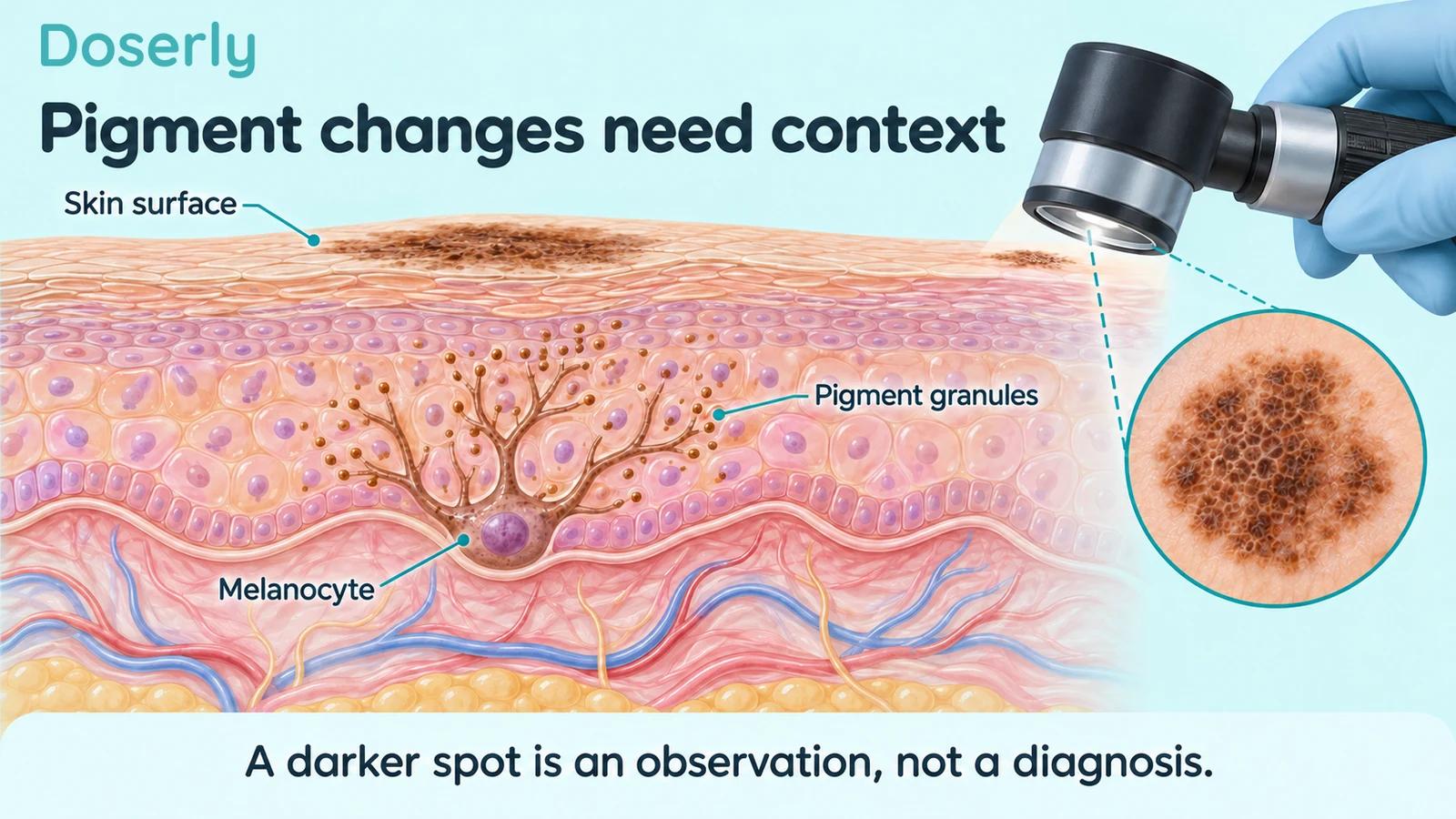
Afamelanotide stimulates pigment-related signaling through MC1R, the melanocortin-1 receptor. Increased eumelanin is part of its pharmacological effect. An existing pigmented spot may therefore become more noticeable without that observation, by itself, establishing a new diagnosis. Scenesse label.
Appearance is only one part of a lesion’s story. A clinician may also consider its shape, distribution of pigment, symptoms, evolution and relationship to other marks on the person’s skin. Treatment dates can help explain when a change occurred, but timing alone cannot determine its cause.
The useful distinction is between recognizing an expected general effect and deciding what a particular lesion means. The first can be described in product information. The second requires assessment of the individual finding.
A small dermoscopy study offers a concrete example
A 2021 study followed 15 adults with EPP, examining acquired melanocytic nevi before and after afamelanotide treatment. Researchers observed pigment-network and globule changes that later moved back toward baseline. They did not observe dermoscopic features suggestive of malignancy during the study. Arisi and colleagues.
That finding helps explain why “darker” and “malignant” are not synonyms. It also shows the value of recording a baseline and comparing structured examinations over time.
The study cannot establish that every changing mole during treatment is harmless. It involved few patients and limited follow-up, and the number of lesions examined is not the same as the number of independent people studied. Its useful contribution is a documented pattern under observation, not a rule that overrides an individual clinician’s concern.
Why the recommendation covers the whole body
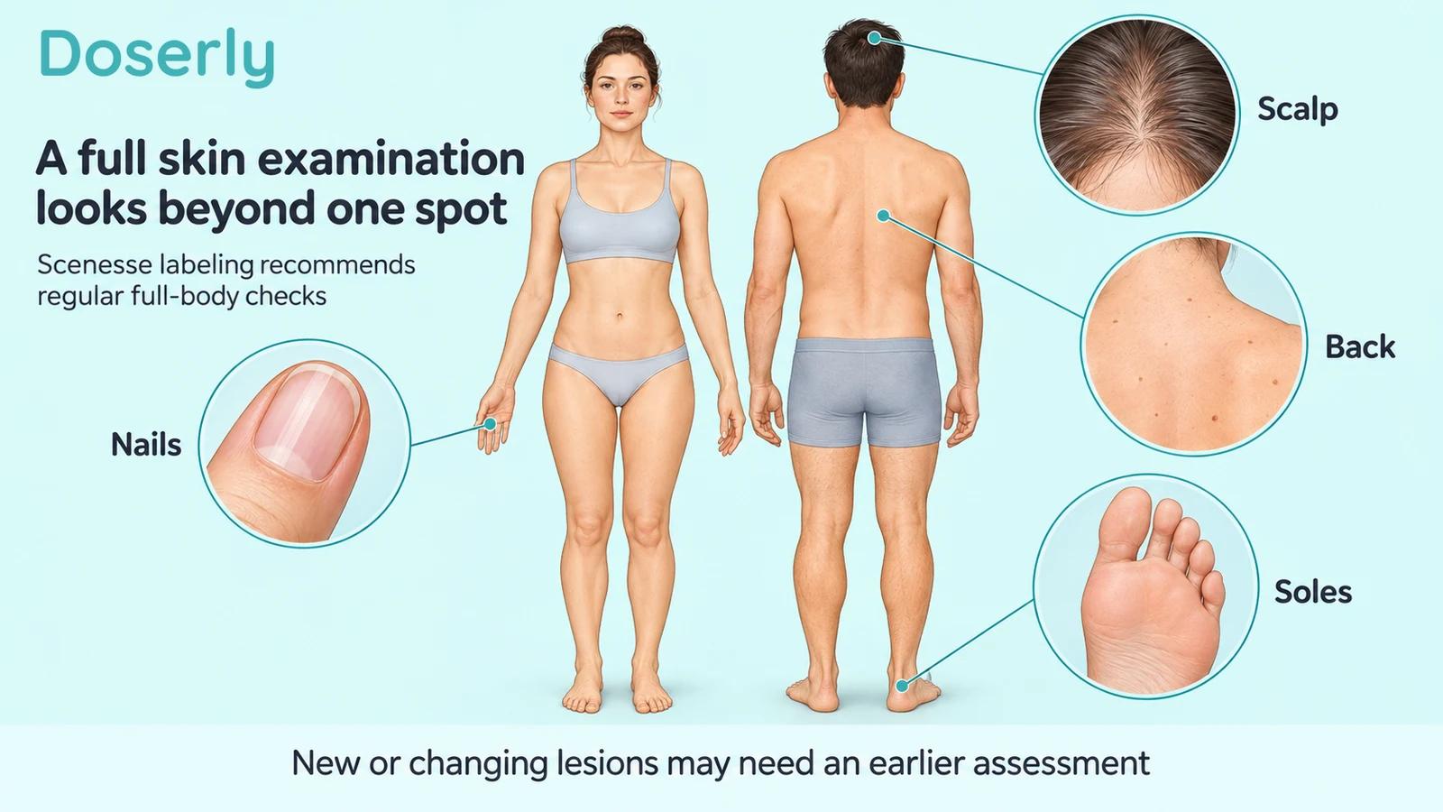
A treatment-related darkening effect may draw attention to one familiar freckle or mole. A full-body examination has a broader purpose: to assess other pigmented lesions and skin abnormalities, including areas a person rarely sees.
European information also notes that people whose EPP improves may increase their sunlight and UV exposure. Monitoring therefore addresses both changes in pigmented lesions and the broader skin context as activity changes. European product information.
This is why an implant-site check alone does not fulfill the same purpose. A local check asks how the administration area is doing. A full skin examination asks about the rest of the skin as well. Both can be relevant, but they are different observations.
A scheduled check is not a reason to wait
A six-month monitoring schedule describes routine follow-up. It does not mean a new concern should be held until the calendar reaches the next appointment.
The European patient information advises reporting new or changing abnormalities and arranging an earlier appointment for growing pigmented lesions or other concerning changes. General dermatology guidance likewise recommends assessment of a spot that changes, differs from others, itches or bleeds. European leaflet, AAD skin-check guidance.
When contacting the clinic, describe what changed and over what period. “A mole is darker” provides less information than its location, whether its outline or size also changed, when it was first noticed and whether there are symptoms. You do not need to decide what the diagnosis is before asking for an appointment.
The ABCDE reminder helps notice changes, not diagnose them
The National Cancer Institute describes the familiar ABCDE features: asymmetry, an irregular border, uneven color, diameter and evolution. These are prompts for noticing a possible concern. Melanomas can be smaller than the often-mentioned six-millimeter size, so a small spot is not automatically exempt from assessment. NCI melanoma appearance guide.
The reminder should not become a scorecard that gives permission to ignore a lesion unless it meets every item. A spot can deserve attention because it is new, changing or unlike the person’s other marks. Skin cancer can also appear in forms other than a dark mole, including a persistent growth or nonhealing sore. AAD skin-cancer overview.
For someone using a pigment-stimulating medicine, “the drug darkens moles” is therefore relevant background, not a diagnostic shortcut. The change still needs to be interpreted in the setting of the whole finding.
Establishing a useful baseline
Before treatment, discuss how the treating team will document the skin and arrange ongoing examinations. Bring information about prior skin cancers, biopsies, atypical moles and family history. European product information calls for special caution with relevant personal or family melanoma history and certain other malignant or premalignant skin conditions. European product information.
A baseline does not certify that no future problem can occur. Its value is comparative: it helps the clinician distinguish a longstanding feature from something newly noticed or changing.
When dates are uncertain, say so. A mark first noticed after an implant may have existed earlier without attracting attention. Separating the date it was noticed from the date it actually appeared helps avoid a misleading causal timeline.
If treatment has already started and no baseline photograph exists, that is not a reason to postpone care. A clinician can assess the current skin and establish a useful record from that point. An imperfect history is still useful when its uncertainties are stated honestly.
How to make photographs useful
If the dermatologist asks for photographs, use their instructions. Clear focus, consistent framing and unfiltered color make comparisons more informative. An overview can show the body location, while a closer image can show the feature of concern. The AAD also recommends checking for blur, glare and shadows and using the clinic’s preferred secure method to send images. AAD photography guidance.
For someone with EPP, obtaining a photograph should not require painful light exposure. Ask the clinic for an approach compatible with your photosensitivity rather than following generic advice to stand in bright sunlight.
A photograph records appearance under particular conditions. It cannot feel a lesion, provide every dermoscopic detail or establish a tissue diagnosis. Camera processing can also change apparent color, which is one reason to avoid judging a subtle difference from two unrelated photographs.
What happens when a clinician is uncertain?
Uncertainty is not a failure of the examination. It can be the reason to use a closer assessment, compare earlier records or obtain a biopsy when appropriate.
A dermatologist may examine a lesion with a dermatoscope, which helps assess structures not readily visible to the unaided eye. In the afamelanotide study, this allowed researchers to describe pigment networks and globules rather than simply calling every lesion darker. Dermoscopy study.
When skin cancer is suspected, examination and tissue assessment serve different roles. The clinical examination identifies what warrants investigation; a biopsy can provide tissue for diagnosis. The AAD’s overview explains that a dermatologist may remove a sample for microscopic evaluation when a spot is suspicious. Skin-cancer diagnosis information.
The decision depends on the lesion and person. An online description of what happened to someone else cannot determine whether your mark should be observed, photographed or biopsied.
Monitoring does not prove that the medicine causes cancer
A monitoring recommendation can exist because a treatment changes what clinicians observe, because a potential risk requires surveillance, or because the consequences of missing a problem are important. It is not automatically evidence that the medicine has been shown to cause the monitored condition.
The FDA’s 2019 Scenesse risk review did not identify an increased melanoma signal in the data it assessed, while still supporting skin monitoring and longer-term safety collection. That is a historical assessment with a defined evidence base, not proof of zero future risk. FDA review.
A 2025 German observational study reported safety and effectiveness information from 200 treated patients. Such follow-up helps characterize experience during clinical use, but this cohort had no untreated comparator and manufacturer involvement. It cannot establish that all rare outcomes have been excluded. German postauthorization cohort.
Nor should a case report be dismissed
A 2026 case report described multiple melanomas in situ in a patient with tanning-bed use, exposure to Melanotan II and I, and other risk factors. The report could not isolate a causal contribution from each exposure. Original case report.
Its practical lesson is not that every darkened mole is cancer. It is that a change associated in time with a tanning product still deserves assessment. A plausible explanation for darkening does not establish that the lesion underneath is benign.
The approved implant and an online tanning preparation also should not be treated as equivalent exposures. Product identity, formulation and monitoring differ. If an unapproved product was used, tell the clinician its name, source and approximate timing rather than assuming the general word “melanotan” provides enough detail.
Keep routine observation manageable
Skin awareness does not require examining every mark repeatedly throughout the day. Ask the treating team how often self-checks are appropriate and what changes should prompt contact. A consistent, manageable approach is more useful than frequent photographs taken under changing conditions.
The AAD’s self-exam guidance includes areas people can miss, such as the back, scalp, soles, between the toes and nails. A mirror or help from someone trusted may make those areas easier to inspect. AAD self-examination guide.
If you find something concerning, record it and contact the clinic. Do not spend weeks trying to prove that it matches an internet example. The purpose of self-observation is to bring a possible change to professional attention, not to replace that assessment.
The rest of treatment follow-up still matters
Skin examinations address one part of afamelanotide care. They do not replace discussion of treatment benefit, implant-site symptoms or other adverse effects. The current US label also includes serious hypersensitivity, including anaphylaxis, which requires a different response from routine pigment monitoring. US prescribing information.
An old safety review should therefore not be used as the sole source for current precautions. Labels can change as more experience becomes available, and the treating team should use the current product information.
At a routine visit, it can help to keep the questions distinct: what benefit has treatment provided, what skin changes have occurred, what adverse symptoms need review, and when should follow-up happen? This makes it less likely that an expected tan overshadows a separate concern.
The Doserly Melanotan I guide provides the wider formulation and evidence context. The practical principle is straightforward: document relevant changes, keep the recommended examinations, and ask for earlier assessment when something new or evolving needs attention.
Sources
- Current US Scenesse prescribing information.
- Current European product information and patient leaflet.
- Arisi et al.: clinical and dermoscopic nevus changes.
- AAD: skin self-examination.
- NCI: melanoma appearance and ABCDE features.
- AAD: skin-cancer symptoms and diagnosis.
- AAD: photographs for a dermatologist.
- FDA: 2019 Scenesse risk review.
- Homey et al.: German postauthorization cohort.
- Vadner and Smith: 2026 melanoma case report.