
Red Light Therapy
October 17, 2026
Red light therapy for scars and wound healing: looking beyond before-and-after photos
On this page
A healing incision can change almost every week. The redness softens, the edges settle and a firm line gradually becomes less noticeable. If someone starts red light during that period, a later photograph may look convincing even when it cannot tell us how much the light contributed.
Human trials give a more useful, and more mixed, answer. Some have found better scar ratings or less postoperative discomfort with particular light treatments. Others have found no meaningful advantage over sham treatment, including studies designed specifically to test skin healing. Recent research also shows how an early improvement on one measure can coexist with no difference on another.
Red light is therefore best understood as a possible addition to selected clinical care, with evidence that depends on the wound, device and outcome. It is not an established way to erase any scar, close every wound faster or replace attention to an incision that is deteriorating.
Wound closure and scar quality are different jobs
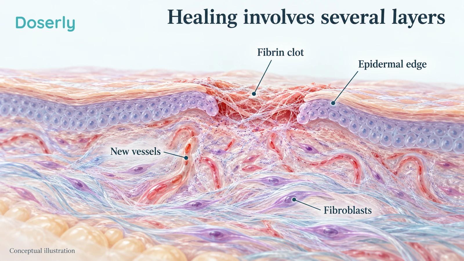
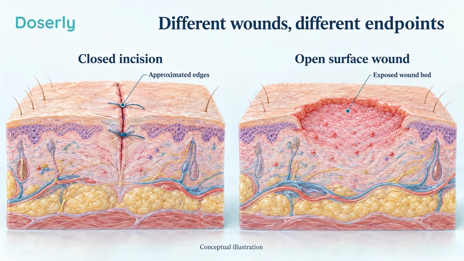
The first task of a wound is to restore a protective surface. An incision whose edges have been brought together has a different job from a broad open area that needs new surface coverage. Below that surface, repair continues as connective tissue is laid down and reorganized.
The American Academy of Dermatology explains that injury depth helps determine scarring. Superficial injury and damage extending into deeper skin do not leave the same repair problem. A closed wound can still have a scar that is red, firm, tender or raised, and those qualities may change over time.
This distinction is easy to lose in the phrase “faster healing.” A paper might use that phrase for a better scar-rating score, less redness, a smaller open area or more complete epithelial coverage. Those findings may each be useful, but they answer different questions. None automatically means fewer infections, greater tissue strength or an invisible final scar.
For a person recovering from surgery, the meaningful outcome may be less pain when getting dressed. For someone with an established scar, it may be less tightness near a joint. Someone else mainly wants a less visible line. Naming the goal makes the research much easier to interpret.
More collagen is not always the desired outcome
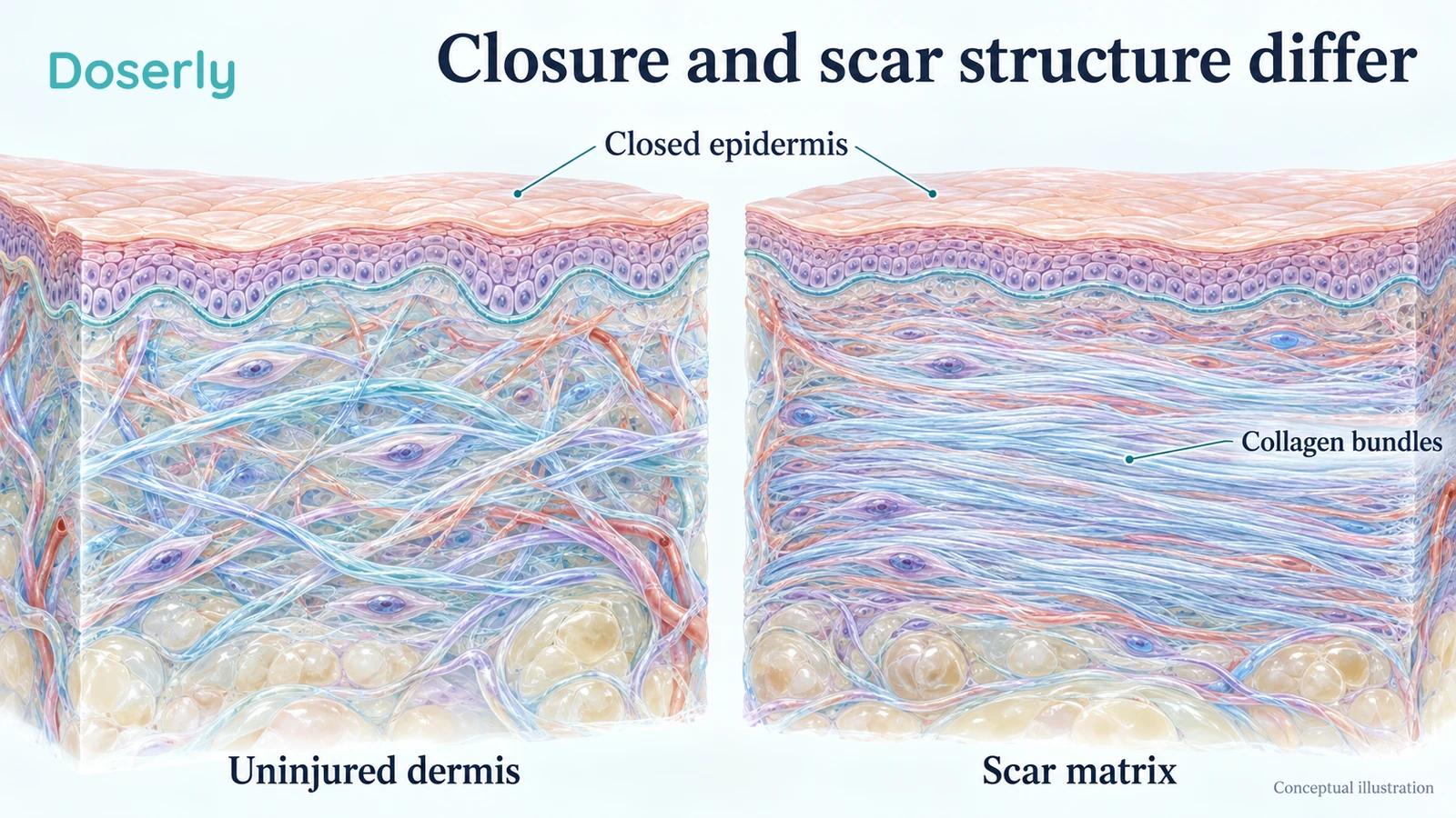
Light treatments are often advertised as stimulating collagen. That can sound appealing in discussions about wrinkles, but scar care requires a more careful idea of what improvement means. A raised, stiff scar already contains an altered connective-tissue structure. Simply making more material is not the same as making that tissue softer or better organized.
The AAD distinguishes flat, raised, depressed and other scar presentations. Their appearance depends on factors including wound depth, location and the person's healing response. A thick raised scar is a different target from a shallow depression after acne. A scar that restricts movement deserves attention to function as well as appearance.
This helps explain why researchers measure flexibility, thickness and symptoms rather than assuming that a collagen-related laboratory change is beneficial. A cellular response may suggest a mechanism worth testing. The patient still needs an outcome that matters in the right direction.
It also explains why early postoperative treatment cannot automatically be treated as evidence for an old scar. Preventing an unfavorable repair pattern while a wound is evolving is a different challenge from changing tissue that has been established for years. If a device advertisement moves between those claims, look for separate human evidence supporting each one.
A direct red-LED scar trial had a negative primary result
The CURES trial, published in 2021, is especially relevant because it tested red LEDs on surgical scars rather than borrowing evidence from an unrelated condition. Thirty women undergoing facelift surgery received light on one side and temperature-matched mock treatment on the other. Treatment began about a week after surgery, with three energy groups.
The primary outcome was scar pliability, a measure related to how firm or flexible the tissue was. There was no statistically significant difference between the treated and control scars on that primary comparison. Some other ratings and changes over time favored particular treatment groups, but they should not be presented as if the main trial result were clearly positive.
Only twenty participants completed all nine sessions per protocol. Treatment-site events included two cases of blistering and one of swelling; they resolved without lasting consequences. These were supervised research exposures, not a template for home dosing. Two investigators disclosed advisory roles with a medical-technology company, and one also disclosed consultancy.
The trial gives us both useful clinical information and a boundary: promising secondary signals do not establish reliable prevention of surgical scarring, and “LED” does not mean adverse effects are impossible.
Near-infrared thyroid-surgery research was more encouraging
A 2022 randomized, double-blind, sham-controlled trial tested an 830 nm LED device after thyroidectomy. Forty-three participants completed it, with twenty-one receiving active treatment and twenty-two receiving sham treatment. Treatment began one week after surgery and continued for four weeks, with follow-up through six months.
At six months, the active group had better satisfaction and clinical scar ratings and lower reported pain. However, the between-group differences on the three-dimensional imaging measurements, including height and color-related features, were not statistically significant.
That combination is more informative than a simple “works” label. It suggests a possible benefit in how the scars were judged and experienced, while objective imaging did not confirm every proposed improvement. It also concerns a specific postoperative neck incision and near-infrared device. It does not validate shining an arbitrary red panel on the thyroid or treating thyroid disease.
A device used at home within a surgical trial is still part of a selected clinical protocol. The wound, timing, instructions and follow-up are part of the intervention, not incidental details that can be discarded when choosing a different product.
An early improvement may not remain a year later
A small abdominoplasty study followed seventeen women whose scars were divided into treated and untreated sides. The right side always received the multiwavelength light treatment, so the study was controlled within each person but not randomized by side.
Scar ratings favored the treated side during the first six months. At one year, participants did not notice a difference between sides. The study included only White women, which further limits generalization across different populations and healing patterns.
This is a useful example of why timing matters. A temporary improvement could still be valuable if it reduces distress or discomfort during recovery. It is simply a different claim from permanently improving the final scar. Without later follow-up, an early photograph cannot tell those possibilities apart.
When discussing treatment, ask whether the proposed benefit is earlier settling, a better eventual result or both. If the evidence only concerns the first few months, that should be part of the decision rather than something discovered after buying a long course of sessions.
Studies after laser resurfacing have also found no clear benefit
Controlled superficial injury can help researchers compare healing within the same person. It does not reproduce every surgical wound, but it is a useful way to test whether a light exposure changes recovery under reasonably similar conditions.
In the HeaLED study, ten healthy volunteers had small forearm areas treated with fractional ablative laser. Different LED wavelengths and intensities were compared with placebo, with measurements including redness and water loss. The primary redness comparison at seventy-two hours was not significantly different, and neither were the other studied measures. The paper appeared online in 2021 and in a 2022 journal issue.
A 2024 study in twenty-five volunteers tested a commercially available combination of blue, red and near-infrared LEDs after fractional resurfacing on the upper arms. It likewise did not find a statistically significant difference in healing. The investigators disclosed research support and equipment from the device company.
These studies do not prove that every possible postoperative LED approach is ineffective. They do show why “light speeds healing after procedures” is too broad. Different wavelengths and combinations have been tested without demonstrating the hoped-for advantage, even when the biological rationale sounded plausible.
If a clinic offers an optional LED session after a procedure, a useful question is whether it has evidence for that particular procedure and outcome. A comfortable experience may be welcome, but comfort during a session is not proof of faster tissue recovery.
What the newer postpartum trials add
Postpartum wound studies are relevant to this question because they separate pain, early wound appearance and medication use. They also show why one positive result should not be extended to every form of childbirth-related injury.
A June 2026 randomized cesarean-section trial included 104 women. Compared with standard care, the light group had about a 1.4-point lower pain score at forty-eight hours and better early scar-scale ratings. Extra analgesic use and global health perception did not differ significantly. Twenty-three participants missed the second assessment, largely because of hospital discharge; their data were handled through imputation.
The comparison did not use a sham device. More importantly, a scar scale measured around forty-eight hours cannot establish the final scar months later. The paper also calls its light infrared while specifying 660 nm, a visible-red wavelength, in the methods. That reporting inconsistency is another reason not to turn the publication into a reproducible home protocol.
Other recent trials have been less positive. A two-center trial in sixty women, published online in late 2025, compared a single red/infrared treatment with sham treatment after perineal trauma. Pain improved in both groups, without significant between-group advantages in pain or healing. High satisfaction did not change that result.
In a separate June 2026 trial of fifty-nine women, red-light treatment after perineal lacerations or episiotomy did not improve the main pain or healing comparisons versus placebo. The active group used fewer analgesics and had longer intervals between doses. That secondary finding is worth reporting without recasting the overall trial as a clear healing success.
An earlier sham-controlled episiotomy trial in fifty-four women also found no significant healing benefit. Taken together, the trials do not justify a universal postpartum light routine. They support a more specific discussion with the team managing the injury, rather than treating a surgical incision and a perineal wound as interchangeable.
An open wound may show mixed changes too
A 2025 randomized trial of thirty patients studied donor sites after a gum graft was harvested from the palate. Both groups received a fibrin sponge; one also received dual-wavelength photobiomodulation.
More treated sites had complete epithelial coverage by day ten. Yet the investigators did not find significant improvements in overall wound closure, pain or analgesic consumption. This is not necessarily a contradiction. The study examined several ways of describing recovery, and they did not all move together.
The location also matters. A palatal donor wound is oral tissue cared for in a dental setting. It cannot establish that a consumer face mask heals a skin abrasion or that a body panel improves a chronic leg wound. Its value here is the lesson about outcomes: an early surface-coverage signal should remain that specific finding.
For an open wound, the priority is a plan that addresses the reason it is open and the factors preventing progress. A photograph of a smaller area can be useful information, but it does not establish that the wound is fully closed, free of infection or ready for normal loading and activity.
Clinical scar lasers are not the same as red-light panels
Some scar treatments deliberately create controlled thermal effects or target blood vessels. These include pulsed-dye and fractional lasers. They should not be grouped together with low intensity photobiomodulation simply because all use light.
A 2022 randomized trial of combined pulsed-dye and fractional nonablative laser compared treatment with healing over time without laser treatment. Of seventy-six participants, fifty-two completed the study. At thirty-six weeks, patient and clinician scar ratings favored the laser group, including improvements in features such as stiffness, thickness or vascularity.
That is evidence for the clinical laser combination studied, with meaningful attrition to keep in mind. It is not evidence that a red LED panel produces the same tissue remodeling. Different devices can use different biological effects, and they require different expectations, training and precautions.
This distinction also works in the other direction. A negative LED trial does not mean every professionally delivered scar procedure lacks value. The useful question is which treatment has evidence for the kind of scar and the problem you actually have.
Start with wound care and the type of scar
After surgery, your own team's instructions take priority over a generic device routine. Those instructions account for the closure, dressing, procedure and any specific concern about healing. Adding light should not mean repeatedly removing a dressing that is meant to stay in place or changing wound care to fit a device session.
For minor skin injuries, AAD wound-care guidance emphasizes cleaning, maintaining an appropriate moist environment, protection and sun care after healing. These ordinary steps have a direct role in recovery. They should not become an afterthought while attention shifts to an optional technology.
Once the wound is closed, the discussion changes. The AAD describes scar treatments that may include silicone, selected injections, pressure approaches or procedures, depending on the scar. Silicone sheets are used on closed wounds, and they can themselves cause irritation. Treatment choice depends on the diagnosis and tolerability rather than a universal ranking.
A raised scar, a keloid, a depressed scar and a scar tethering movement may need very different plans. If the main issue is tightness across a joint, ask about function and rehabilitation rather than focusing only on color. If the main issue is appearance, it is reasonable to ask what degree of change is realistic and whether time alone may improve it.
Light can be considered within that conversation. It should not require pretending that the basic distinction between an open wound and an established scar has disappeared.
Know when the question is no longer cosmetic
An incision that is becoming more painful, swollen, hot or increasingly red, especially with pus or fever, needs clinical assessment. NHS surgical-wound guidance identifies these as possible signs of infection. A wound separating or otherwise deteriorating should also be reported to the treating team.
Red light is not a way to postpone that assessment. Less discomfort after any intervention would not by itself exclude a complication. Likewise, no amount of confidence in a device specification can tell you whether an unexpected wound problem needs antibiotics, drainage, a dressing change or another intervention.
A sore that repeatedly breaks open or does not heal also deserves diagnosis, particularly if there is no clear explanation. The AAD notes that skin cancer can resemble a persistent sore or scar-like mark. This does not mean every slow-healing spot is cancer; it means the cause should not be assumed from its appearance or treated indefinitely as an ordinary scar.
If a scar follows removal of a skin cancer, discuss any proposed light exposure with the treating clinician. Cosmetic healing goals and the follow-up plan for the original lesion need to be considered together.
What a useful assessment looks like
A good scar assessment asks more than whether the line looks lighter. Clinicians may consider its height, flexibility, color, symptoms and effect on movement. Patient-reported discomfort can matter even when two photographs look similar.
The original Patient and Observer Scar Assessment Scale study deliberately included separate patient and observer scales. Its initial testing involved forty-nine burn-scar areas in twenty people, assessed by four independent observers. This history is a helpful reminder that the person living with a scar has information that an outside observer cannot obtain just by looking. It also reminds us to check the population in which a measurement was tested: a burn scar and a fresh surgical incision are not identical situations.
When a paper reports a better total score, look for the components behind it. A combined score can improve because one feature changes while another remains much the same. That does not make the improvement meaningless, but it affects what someone should expect. A reduction in conspicuous redness may be worthwhile without implying that a tight scar has become more flexible.
Consider two practical examples. A person whose sleeve no longer catches on a raised scar may value a small change that is difficult to capture on a phone. Another person might see a paler line but still experience the same pulling sensation when reaching. Calling both situations “better healing” conceals the difference that matters to each person. Bringing those details to a follow-up visit helps the clinician decide whether the current plan is addressing the right problem.
For the same reason, agree on a review point before adding another treatment. Otherwise, it is easy to keep extending sessions while waiting for an undefined improvement. A clear goal does not guarantee success; it makes success or lack of progress easier to recognize.
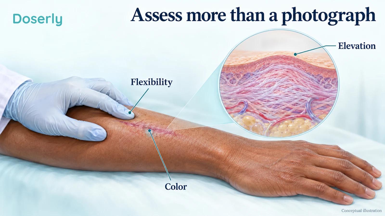
For your own records, note the date of the procedure or injury, when the wound closed and when each new treatment started. If you take photographs, keep the lighting, distance and body position comparable. A relaxed arm and a stretched arm can make the same scar look different. Avoid interpreting ordinary changes in posture, exposure or camera contrast as tissue remodeling.
Also record what you can do more comfortably. Wearing a waistband, reaching overhead or sleeping without the scar rubbing may be more informative than a small change in redness. These are practical observations to discuss at follow-up, not a replacement for examination or a validated home scoring system.
When a clinician proposes photobiomodulation, ask which outcome is expected to improve, what evidence applies to your wound, and when progress will be assessed. Ask whether the aim is early comfort, faster surface recovery or a better mature scar. A plan with those distinctions is easier to evaluate than a promise to “boost healing.”
The research supports continued investigation and some carefully selected clinical use, while leaving substantial uncertainty about everyday home treatment. The most defensible expectation is a possible additional benefit for a particular wound or scar, with ordinary healing, standard care and measured follow-up kept firmly in view.