
Red Light Therapy
October 17, 2026
Red light for childhood myopia: benefits, rebound and retinal safety
On this page
When a child needs a stronger glasses prescription at every visit, a treatment that might slow the process deserves attention. Parents are thinking about more than the next pair of lenses. They want their child to see comfortably now and to have healthy eyes decades from now. Red-light treatment has entered that conversation because several clinical trials have measured substantially less eye growth during treatment.
The difficult part is that the same treatment deliberately exposes the retina to light. Its benefit and its safety therefore need to be considered together. The evidence available through October 2026 includes positive randomized trials, a small Australian study, new reports of retinal changes, device measurements that raise safety concerns, and evidence that progression can accelerate after treatment stops. It supports serious clinical investigation, but does not establish a safe do-it-yourself approach.
For a family weighing the idea, the useful question is whether a particular, appropriately regulated treatment offers a favorable balance for their child compared with other myopia-control options. A promising average result cannot answer that alone. The child's age, documented progression, retinal health, available alternatives, and ability to attend follow-up all belong in the discussion.
What researchers are trying to change
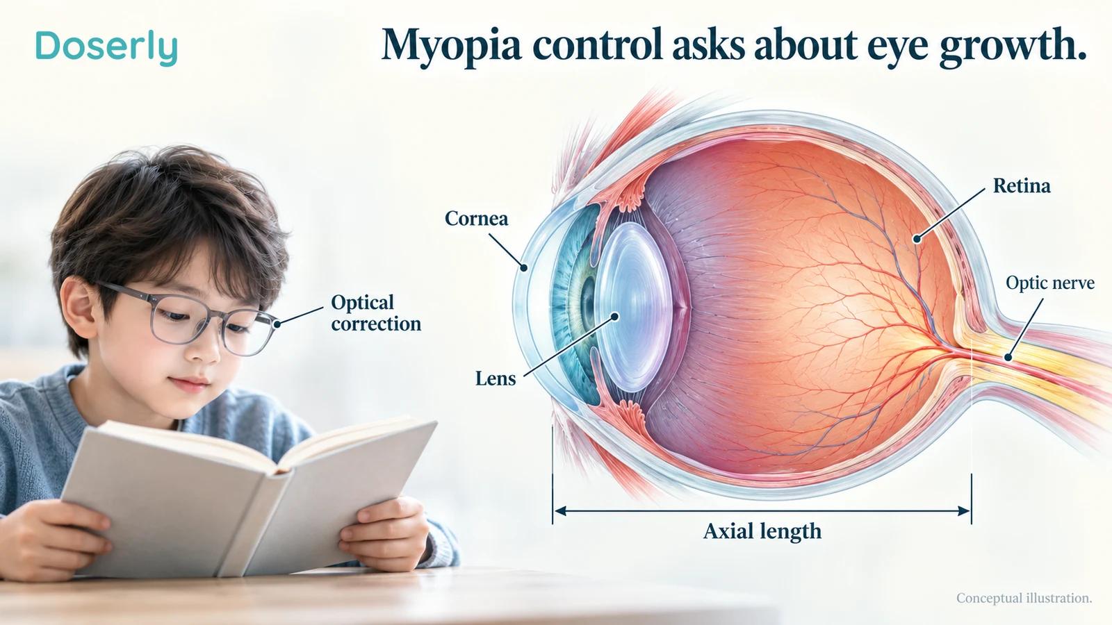
Myopia means that distant objects are blurred because light focuses in front of the retina. Often, the eye has grown too long from front to back, although the cornea and lens also contribute to focusing. Ordinary prescription glasses can bring the image into focus without addressing continued elongation. Severe myopia carries additional risks, including retinal detachment. The National Eye Institute explains the anatomy and usual course.
That distinction explains two measurements that appear repeatedly in trials. Axial length describes the front-to-back length of the eye, usually reported in millimeters. Spherical equivalent refraction summarizes refractive error in diopters. A study can ask whether the eye grew less, whether the prescription became less myopic, or both. Corrected visual acuity, the ability to read an eye chart with appropriate lenses, answers another question.
These are related outcomes, but they are not interchangeable. A child may read clearly through updated glasses while their myopia continues progressing. Conversely, a small measured change in eye length does not establish that the child no longer needs glasses. Keeping those outcomes separate helps families interpret a headline such as “myopia reversal” without mistaking a short-term measurement for a lasting cure.
The measurements also depend on consistent examination. Trials commonly use cycloplegic refraction, which temporarily relaxes the eye's focusing system, alongside optical biometry for axial length. Comparing a casual vision check with a trial's carefully measured endpoint can give a misleading impression of progress.
The randomized evidence shows a real treatment signal
A foundational multicenter trial randomized 264 Chinese children aged eight to thirteen to repeated low-level red-light treatment plus single-vision spectacles or spectacles alone. At twelve months, estimated axial elongation was approximately 0.13 millimeters with treatment and 0.38 millimeters in the comparison group. Refractive progression also favored treatment. The analysis included 246 children with follow-up, and 225 completed the twelve-month visit. The original trial describes the comparison and participant accounting.
This was a substantial difference in eye growth during the study. It was also a comparison against ordinary spectacles, without a sham light device. That design provides stronger evidence than an uncontrolled before-and-after report, while leaving some questions about masking, behavior, and device-specific effects. Its reassuring examination findings over one year cannot establish safety over an entire childhood.
A subsequent double-masked trial randomized 112 children to the treatment device or a reduced-power sham. At six months, average elongation was 0.02 millimeters in the active group and 0.13 millimeters in the sham group; refraction also favored active treatment. Importantly, the sham delivered ten percent of the original power. It was a lower exposure, rather than a completely unilluminated device. The sham-controlled study reports these results.
That result strengthens the case that a treatment effect exists under studied conditions. It does not mean that increasing a device's output will produce more benefit, or that a different device will reproduce the result. Neither inference was tested by comparing the two assigned groups.
What the 2026 studies add
An Australian pilot adds useful information beyond the predominantly Chinese evidence base. It randomized 34 children from multiple ethnic backgrounds to red-light treatment plus spectacles or spectacles alone. At twelve months, adjusted axial-length changes were approximately minus 0.03 millimeters and plus 0.12 millimeters, respectively. Three children in the treatment group withdrew, one because of headache and two because of adherence difficulties. No severe adverse events were documented. The 2026 twelve-month report gives the findings.
The study is encouraging, but seventeen children assigned to each arm is a small foundation for judging uncommon harm or differences across populations. Its earlier three-month publication and the later twelve-month report describe the same pilot, not two independent confirmations. Some investigators had affiliations with the device company, another reason independent replication remains valuable.
A separate 2026 exploratory study tested a noncoherent LED intervention against sham in forty children. Its participant-level analyses did not establish a significant adjusted advantage in axial length or refraction. A nominally significant result in the left eye did not make the overall study definitively positive. Follow-up lasted only three months, and no serious adverse events or structural retinal abnormalities were observed. The original LED trial explicitly states these limitations.
That distinction is especially useful when comparing products. Laser-based treatment and an LED intervention cannot inherit one another's efficacy results because both emit red light. The newer LED study also illustrates why one favorable result among several analyses needs to be read alongside the main participant-level findings.
Changes in the choroid are interesting, but not a cure
Researchers have also measured the choroid, the vascular tissue beneath the retina. In a secondary analysis of 120 children from two sites in the original multicenter trial, red-light treatment was associated with changes in choroidal thickness. Early changes had some ability to predict later myopia-control outcomes. The original analysis identifies its relationship to the parent trial.
This suggests a biological response worth investigating. It does not prove that choroidal thickening is the sole reason treatment works, or that every thickness change is beneficial. The analysis reused participants from an efficacy trial, so it should not be counted as another independent trial showing that treatment controls myopia.
For families, a scan described as “healthier” can sound more definitive than the measurement warrants. Thickness, eye length, retinal structure, and useful vision each contribute different information. An anatomical response becomes clinically persuasive when it fits with durable benefit and acceptable safety, rather than standing in for them.
Why the eye needs a different safety discussion
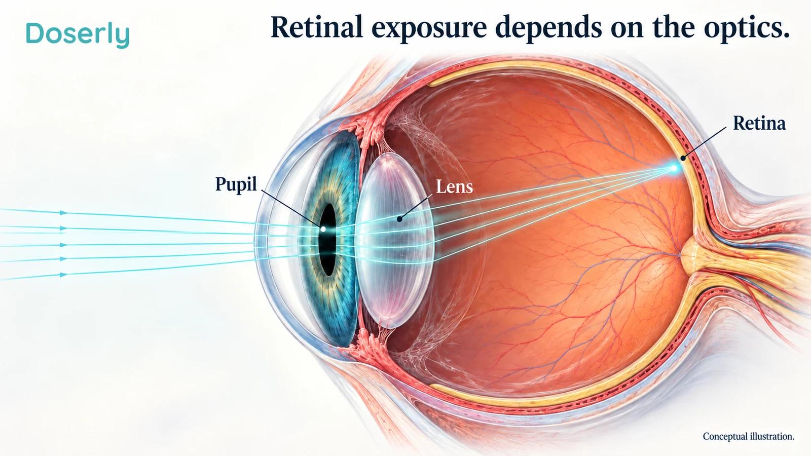
The eye is an optical system. Light entering through the pupil can be concentrated onto a small retinal area. A power figure measured outside the eye therefore does not, by itself, describe the exposure received by retinal tissue. Wavelength matters, but so do the beam, optics, pupil, alignment, and duration.
A 2025 laboratory investigation examined a red-light myopia device and found that retinal exposure could vary with factors including accommodation, pupil size, positioning, and eye movements. Under some tested conditions, exposure exceeded relevant limits. This was a device-measurement study, not a clinical estimate of how often children develop injury. The primary radiometric investigation explains the optical problem.
Its practical implication is not that parents should calculate a new home exposure. It is that a reassuring marketing label cannot replace independent device characterization. A low-looking power number and a comfortable sensation are inadequate substitutes for knowing what reaches the retina.
A 2026 laboratory analysis compared four devices and found substantial differences in their optical safety characteristics. The tested laser devices could reach exposure limits within the manufacturers' treatment periods under the study's conditions, while the tested diffuse LED device had a different safety profile. The JAMA Ophthalmology investigation reports the device-specific assessments.
Those measurements do not establish that every use causes injury, that all laser devices are identical, or that all LEDs are clinically safe. They do show why the safety of one optical design cannot be generalized to another. There is no universal safe session length that can be extracted from these findings for an untested consumer lamp or panel.
Normal vision does not exclude a retinal change
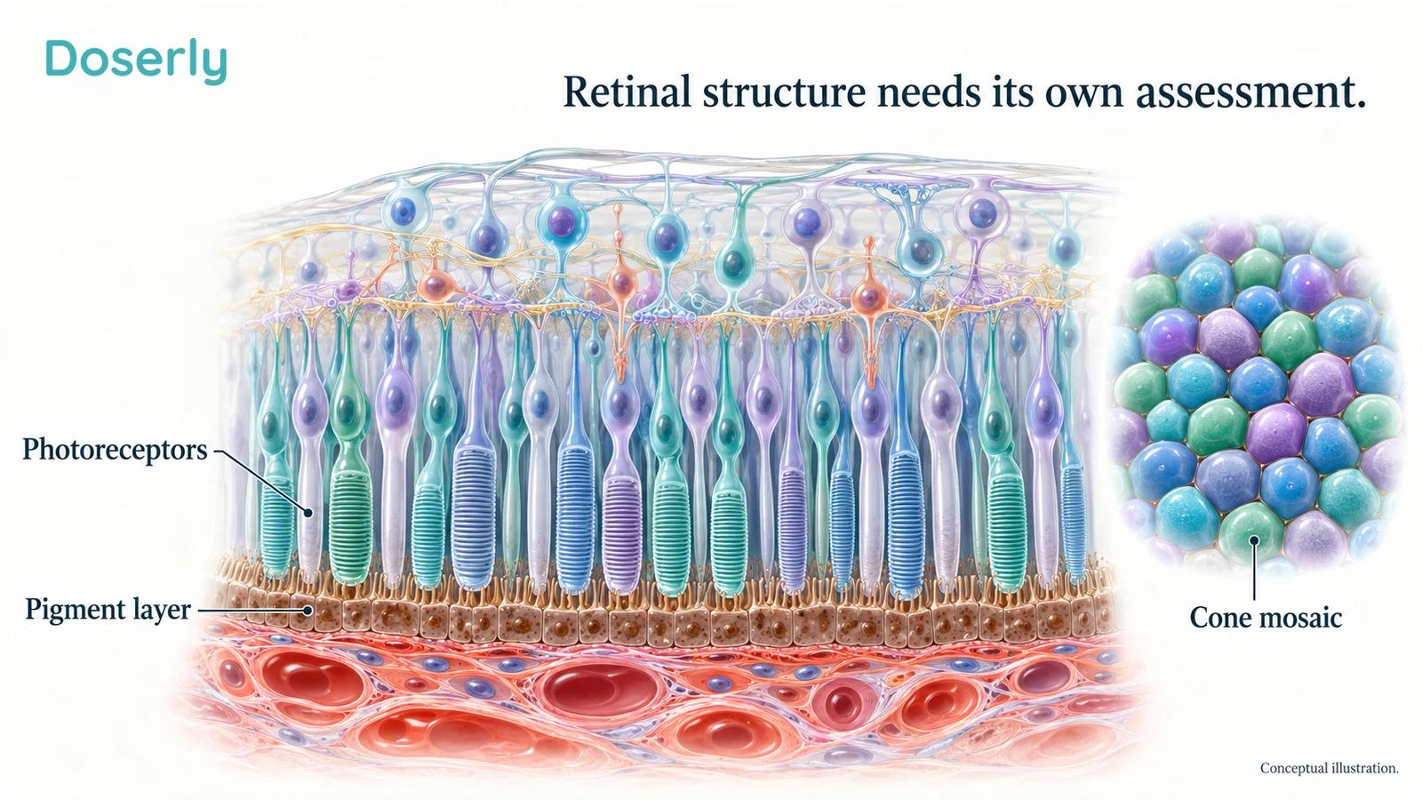
A 2025 randomized study followed 86 children with refractions ranging from mild hyperopia to mild myopia. Three children assigned to red-light treatment developed focal changes on optical coherence tomography, or OCT. They had no reported visual symptoms, and corrected acuity remained unchanged. The changes resolved after treatment stopped, over approximately two weeks to three months. The full original report describes the findings and follow-up.
This study is important precisely because the children did not necessarily complain. OCT examines retinal structure at a level that a symptom report or ordinary eye-chart score may not capture. The study had substantial loss to follow-up and does not provide a universal injury rate for every device. Resolution after stopping is reassuring about those observed events; it does not prove that similar changes are harmless or always reversible.
There is also a published report of retinal damage in a twelve-year-old after repeated low-level red-light exposure. A single case cannot establish the frequency of injury or define all the circumstances that contributed. It does establish that the safety discussion cannot rely solely on trials reporting no serious events. The original case report is indexed in JAMA Ophthalmology.
Different kinds of evidence answer different parts of the question. A case identifies a possible harm. A controlled trial estimates benefit and common short-term events in selected participants. An optical study tests exposure characteristics. None replaces the others, and a favorable result in one category does not erase a concern in another.
What longer follow-up can and cannot reassure us about
A 2026 multicenter real-world study examined 362 current users across treatment-duration groups. Only 91 were in the group treated for at least three years. The investigators reported favorable eye-growth control in that longer-duration group and no treatment-duration pattern in full-field electroretinography, which measures retinal electrical responses. Four eyes had reversible OCT changes. Investigators disclosed related patents and company directorship and shareholding. The original study explains how participants were selected.
It would be misleading to describe this as 362 children all followed for three years in a randomized safety trial. The study sampled people still using treatment. Children who stopped because of difficulty, dissatisfaction, or a problem may be represented differently or missed. The data add useful experience, while that selection limits the reassurance they can provide.
Another 2026 study used adaptive optics to examine the cone photoreceptor mosaic. It compared twenty treated eyes with untreated myopic and nonmyopic eyes and did not find a significant reduction in cone density relative to untreated myopic eyes. This is reassuring for the measurements obtained. However, it was a small cross-sectional comparison, without pretreatment measurements establishing each eye's trajectory. The original retinal-imaging study reports the design.
Together, these reports make the safety picture more detailed, rather than settled. A normal specialized measurement in a small sample cannot exclude uncommon injury, delayed effects, or a problem confined to another retinal structure. Parents deserve to hear both the reassuring observations and the limits of the follow-up.
Rebound matters because childhood treatment eventually changes
A treatment plan needs an ending, or at least a transition. Myopia may progress faster after an intervention stops, potentially reducing some of the advantage accumulated during treatment. That possibility matters when a family is considering years of expense, appointments, and daily participation.
The two-year follow-up of the original multicenter red-light trial found faster second-year elongation among children who stopped treatment than among those who continued with spectacles alone throughout. The corresponding average changes were approximately 0.42 and 0.28 millimeters. However, second-year treatment choices were not randomized, participation fell substantially, and only eleven analyzed children continued red-light treatment. The full follow-up describes the selection and rebound findings.
This is evidence of a concern, not a precise prediction for an individual child. It also does not mean that every earlier benefit was necessarily erased. Both the rate after stopping and the total accumulated change are needed to understand the longer trajectory.
A 2026 randomized comparison adds more direct evidence. It assigned 136 children to red light or one-percent atropine, a high concentration, followed by a three-month washout. Red light produced less elongation during the treatment year, but axial growth during washout was greater: approximately 0.163 versus 0.115 millimeters. Follow-up participation was uneven between groups. The original trial reports both treatment and withdrawal phases.
The comparator concentration matters; this was not a comparison against every low-concentration atropine approach. The authors discussed reduced dosing or tapering as possible future strategies, but the study did not validate a tapering schedule. Families should not improvise one from that suggestion. The evidence supports discussing withdrawal and monitoring before treatment begins, including what would trigger a change in plan.
Other myopia-control options deserve an equally concrete discussion
Red light is entering a field with existing treatment research. Considering alternatives is not an admission that progression does not matter. It is how a family weighs the same goal against different burdens and uncertainties.
The BLINK randomized trial enrolled 294 children and compared soft multifocal contact lenses with single-vision contact lenses. Over three years, the higher-add multifocal design produced less refractive progression and less eye growth. Average axial elongation was approximately 0.42 millimeters with that design and 0.66 millimeters with single-vision lenses. The original trial provides the controlled comparison.
A later BLINK2 follow-up examined discontinuation in older teenagers. Eye growth increased slightly after switching away from multifocal lenses, but the investigators found no evidence that the previously accumulated treatment advantage was lost. The follow-up involved older participants and a different intervention, so it cannot predict what happens when a younger child stops red light. The 2025 primary follow-up explains the findings.
Prescription spectacle options have also advanced. In September 2025, the FDA authorized Essilor Stellest lenses for specified children starting treatment at ages six to twelve, with defined refractive limits and otherwise non-diseased eyes. These are purpose-designed myopia-control spectacles, not ordinary single-vision glasses. The FDA authorization letter gives the exact indication.
Atropine has its own dose-dependent evidence. The LAMP trial randomized 438 children to several low concentrations or placebo and measured refraction, axial growth, pupil size, and accommodation. Benefits and physiological effects differed across concentrations. The original LAMP report shows why “atropine” is not one uniform comparison.
These studies were not a single head-to-head contest against red light. Differences in age, baseline progression, follow-up, and comparison groups prevent a fair ranking by simply placing percentages side by side. The clinician's task is to match an option to the child and explain its practical tradeoffs, including contact-lens handling, visual comfort, medication effects, and follow-up requirements.
Outdoor time helps answer a different question
A large school-based randomized trial in Guangzhou found that adding outdoor activity reduced the incidence of new myopia over three years. Among eligible children without myopia at baseline, the incidence was 30.4 percent in the intervention schools and 39.5 percent in the comparison schools. The axial-length difference across the study population was not statistically significant. The original trial separates these outcomes.
The distinction is useful: preventing or delaying myopia onset is not identical to treating established, progressing myopia. Outdoor activity can be part of a child's healthy routine without being presented as a substitute for prescribed correction or a complete treatment for progression. It also does not mean looking directly at the sun or reproducing a bright-light exposure indoors.
A family should not have to choose between ordinary supportive habits and professional care. The two can coexist. The question is what additional intervention, if any, is justified by the child's measured course.
A useful appointment starts with the child's own trajectory
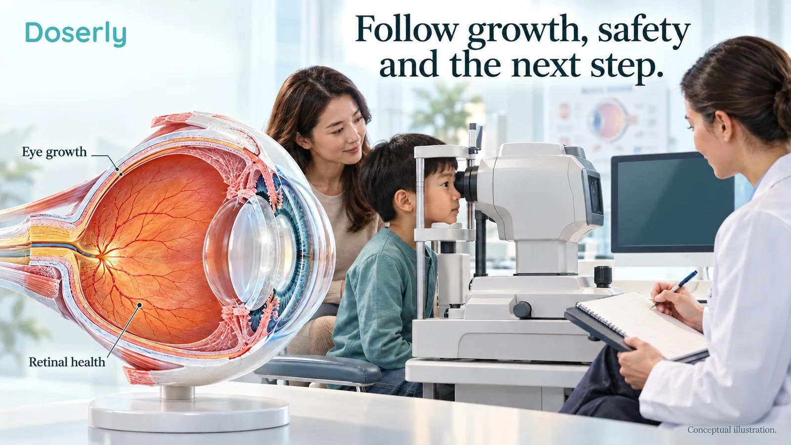
Bring previous prescriptions and examination records if they are available. Ask whether progression has been documented with comparable measurements and whether axial length has been tracked. A single number is less informative than the pattern, especially when different clinics or methods have been involved.
If red-light treatment is proposed, the discussion should identify the exact device, its authorization in the relevant jurisdiction, the evidence for that model, and the clinician responsible for safety monitoring. Ask what retinal assessment is planned, how a change would be recognized, and how treatment would be stopped or changed if necessary. These are part of evaluating a medical intervention, not technical details a parent should have to solve alone.
An eye-treatment authorization for another disease is not transferable. For example, the FDA's Valeda indication concerns selected people with dry age-related macular degeneration and specified visual-acuity and retinal criteria. It is not an authorization to treat childhood myopia with that device, a red lamp, or a consumer panel. The original FDA order defines the adult retinal-disease indication.
For any child, sudden flashes, a marked increase in floaters, or a curtain-like shadow require urgent eye assessment rather than waiting for a routine follow-up. The National Eye Institute identifies these as retinal-detachment warning symptoms. New persistent visual disturbance after an exposure also deserves prompt contact with the treating eye professional; repeating exposure to see whether it happens again is not a useful test.
Red-light myopia research has moved beyond a speculative mechanism: several trials show reduced eye growth during treatment. The remaining decision is still demanding because a child's retina must remain healthy while that benefit is pursued. The strongest conversation holds efficacy, device-specific safety, alternatives, and the eventual transition off treatment in view at the same time.