
Red Light Therapy
October 17, 2026
Red light therapy and pigmentation: melasma, dark marks and skin tone
On this page
A light mask can seem like a gentle answer to stubborn discoloration. It does not peel the skin or visibly remove a dark spot, and many devices promise a more even complexion. But pigment is one area where “light therapy” is too broad a label to be useful. Some wavelengths can provoke pigmentation, some are being investigated as treatments, and some clinic devices deliberately deliver energy in ways a home mask does not.
The evidence does not currently establish a red home mask as a reliable treatment for melasma. There are encouraging small studies of specific photobiomodulation approaches, including amber and near-infrared light. There are also null results, combination trials that cannot isolate the effect of light, and professional laser studies that should not be used to sell ordinary LEDs.
Research available through October 2026 supports a careful distinction between treating a diagnosed pigment disorder and trying to make skin look generally brighter. The most helpful starting point is understanding what the discoloration is, what caused it, and exactly what treatment a study tested.
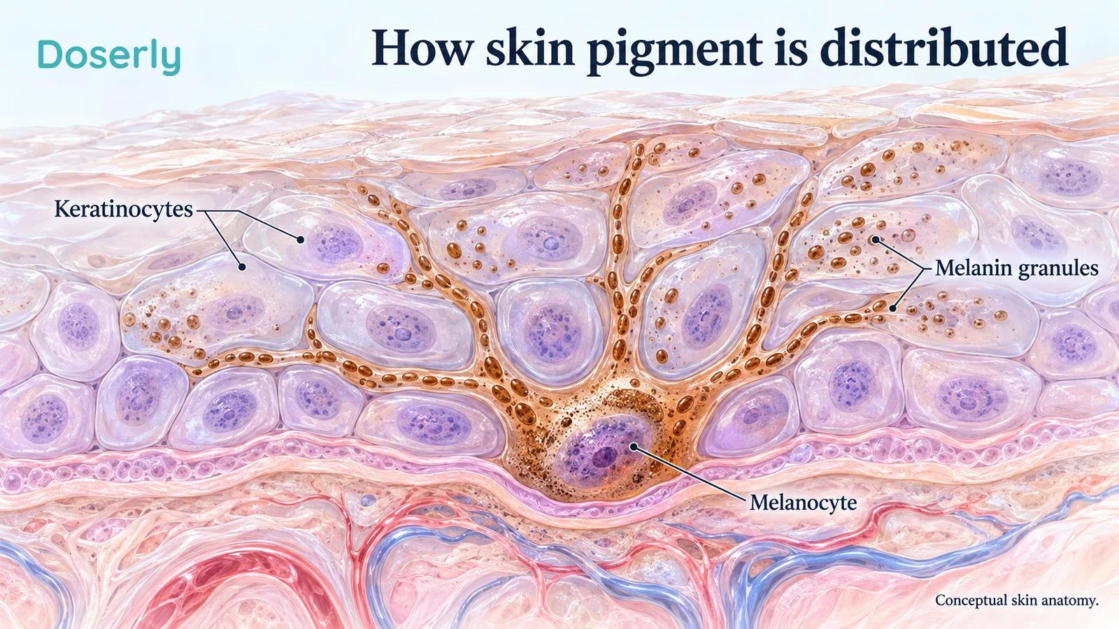
Conceptual anatomy shows pigment distribution within the epidermis. It does not show a light treatment removing melanin or predict a change in skin tone.
Melasma and a mark left by a pimple are different problems
Melasma commonly appears as uneven brown patches across areas such as the cheeks, forehead or upper lip. It tends to be persistent or recurrent. Post-inflammatory hyperpigmentation is the extra pigment left after inflammation or injury, such as acne, dermatitis or an irritating skin-care product. Both can look darker than the surrounding skin, but they do not have identical causes or treatment histories.
The American Academy of Dermatology's guidance on dark spots emphasizes finding and addressing the trigger. If new acne lesions or skin irritation continue, new marks can appear while older ones fade. Treating the source of inflammation is therefore part of pigment care, rather than a separate concern to postpone until after buying a device.
This distinction also changes how improvement should be interpreted. A post-acne mark may gradually fade as the inflammation settles. If someone starts light treatment during that period, the timing alone cannot show how much the device contributed. A study of melasma cannot automatically answer whether that same device speeds the fading of individual acne marks.
It is worth describing the pattern rather than only the color: whether it followed breakouts, appeared during pregnancy, developed after a procedure, or has slowly expanded. An uncertain or changing lesion should be assessed before treating it as a cosmetic dark spot. The goal is a diagnosis that explains the change, not a device setting chosen from a photograph.
Pigment is part of living skin
Melanin is made by melanocytes and distributed in pigment-containing structures called melanosomes to neighbouring keratinocytes. It contributes to normal skin color and protection. Treating an unwanted patch is different from treating someone's natural complexion as a problem.
A biopsy study of fifty-six Korean women with melasma compared affected and nearby skin. Melasma areas contained more epidermal melanin, with more widely distributed melanosomes in keratinocytes and changes in melanocyte activity. The underlying skin also showed more solar elastosis, a change associated with accumulated sun exposure. That picture is more complex than a layer of pigment sitting on the surface waiting to be scrubbed away.
Pigment can also be present deeper in the skin, including inside macrophages called melanophages. The terms epidermal, dermal and mixed describe tissue compartments, not a simple instruction to choose a stronger or deeper-reaching light. Color alone does not reliably tell a person which compartment is involved or which device will help.
For a treatment to be useful, changes in pigment biology need to translate into a meaningful clinical improvement. Reduced activity of an enzyme in cultured cells is interesting. It is not the same as reducing the darkness or area of melasma on a face, preventing recurrence, or avoiding new irritation.
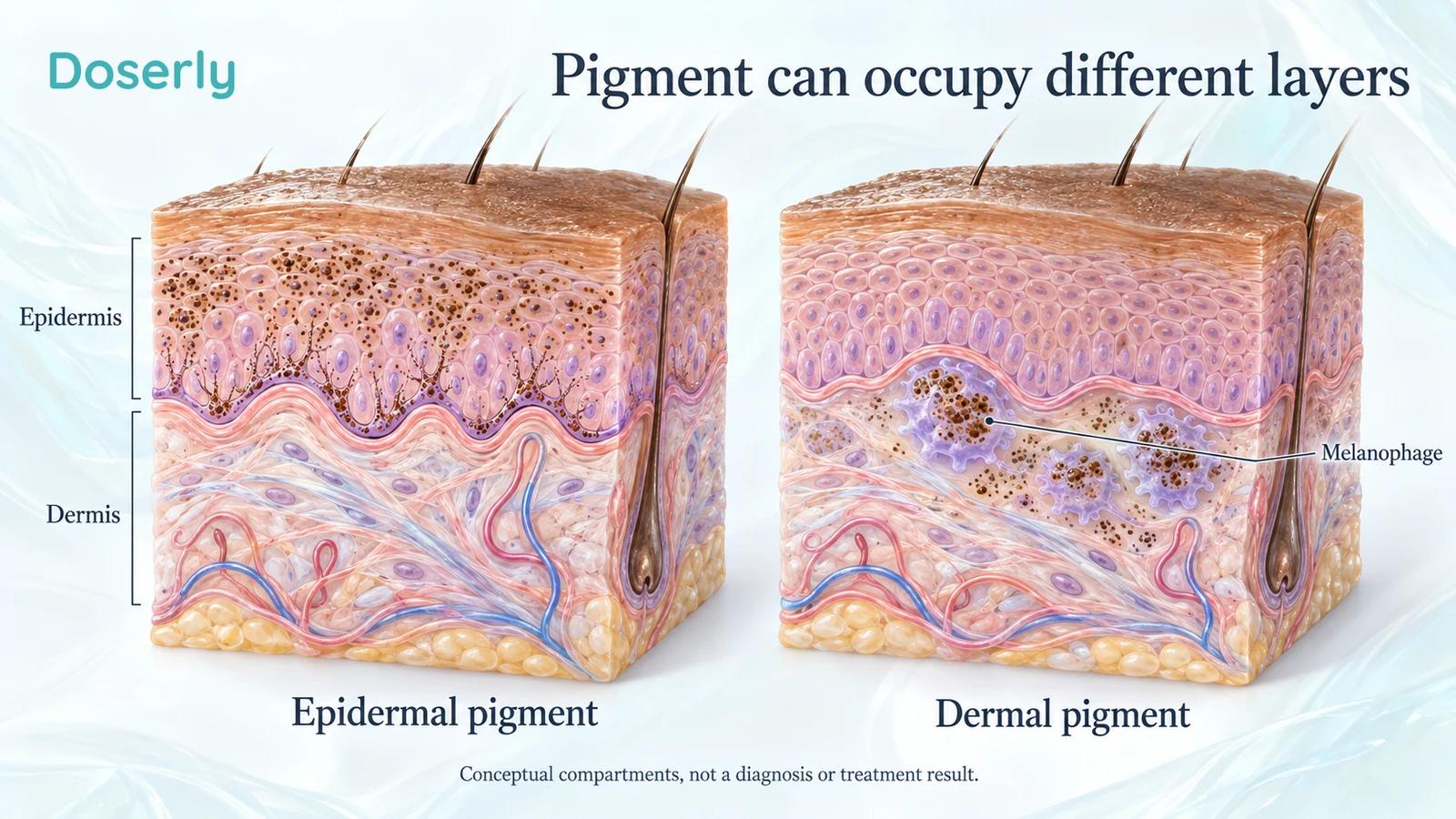
Conceptual pigment compartments are shown separately for clarity. They are not before-and-after images, and an individual's pigment depth requires clinical assessment.
Visible light does not have one effect on pigmentation
People often learn that ultraviolet radiation stimulates pigmentation, then assume visible light is irrelevant. Human exposure studies show that the answer depends on the spectrum and the skin being studied.
In a 2010 experiment involving twenty volunteers with skin types IV to VI, broad visible light produced darker, more persistent pigmentation than the long-wavelength UVA comparison under the tested conditions. The investigators also examined type II skin, where they did not observe the same pigmentation response. This was a controlled exposure study on the back, not a trial of a commercial facial device.
A 2014 wavelength-specific study separated blue-violet light at 415 nanometres from red light at 630 nanometres. In healthy volunteers with skin types III and IV, the blue-violet exposure produced dose-related pigmentation that could persist for months. Red light did not induce hyperpigmentation under those experimental conditions.
These results are useful without turning them into absolutes. They show why it is misleading to describe all visible wavelengths as identical pigment triggers. They do not prove that every red mask is harmless for every person with melasma, or that every blue-light device inevitably causes a dark patch. The spectrum, delivered exposure, skin condition and equipment all matter.
They also do not support equating a screen with a treatment panel solely because both emit visible light. Wavelength is only one part of exposure. A claim about one source needs evidence about that source, rather than borrowing the result of an unrelated laboratory lamp.
The early photobiomodulation studies are promising but small
One 2018 split-face pilot enrolled seven women with dermal melasma. Both sides received microdermabrasion; one randomly selected side also received pulsed 940-nanometre near-infrared light. The light-treated side showed greater improvement in pigment assessments at twelve weeks.
That is an encouraging signal, but it was a seven-person combination-treatment study. Only three participants had follow-up extending to twelve months. It does not establish that red LEDs alone work, that microdermabrasion should be copied at home, or that light can replace sun protection. The paper's suggestion of enhanced resistance to future sunlight remains an exploratory interpretation, not a clinical sunscreen claim.
A 2022 study of amber light combined laboratory experiments with a ten-patient clinical pilot. The researchers investigated vascular-cell activity and reported improvements in facial redness and pigmentation after treatment. The cellular findings and the small clinical observation are different kinds of evidence. Neither establishes a broad effect across skin tones or long-term melasma control.
These studies help explain why researchers continue to investigate photobiomodulation. They are less useful for choosing an arbitrary retail product. If a mask seller cites them, check the wavelength and accompanying treatment. A near-infrared procedure combined with microdermabrasion is not a red-mask trial, and an amber pilot is not proof that adding more colors to a device improves its performance.
A home amber device has been compared with a clinic laser
A randomized study first published in 2023, appearing in a 2024 journal issue, compared home 590-nanometre LEDs with an in-hospital 1064-nanometre Q-switched laser. Fifty participants were assigned to the two groups and assessed over sixteen weeks. Both groups also used sunscreen and received oral vitamins C and E.
Melasma severity scores improved within both groups. The mean reduction was about 2.0 points with LEDs and 3.1 with the laser, but the between-group difference was not statistically significant. This is not proof of equivalence, and the study did not include a group receiving the background care without either light treatment.
The result is relevant because it actually studied home use, rather than merely suggesting it from laboratory data. Its limits are equally relevant: a single centre, a small sample, a particular amber device, and background treatments shared by both groups. It supports further study of that approach, not a claim that any inexpensive home mask performs as well as professional treatment.
For readers weighing convenience against clinical care, the comparison is worth discussing with a dermatologist. It should not become a reason to select a wavelength without first confirming the diagnosis and considering what standard care has already achieved.
The 2025 amber trial did not show a clinical improvement
A 2025 randomized pilot compared active amber photobiomodulation plus placebo cream with sham light plus topical tranexamic acid. Twenty-one women participated over twelve weeks. The primary outcome was the Melasma Area and Severity Index, or MASI.
The study found no significant change in MASI or the physician's global assessment, either within or between groups. Quality of life improved in both groups. The trial had ended early and was underpowered, so it could not establish the intended noninferiority conclusion.
The authors proposed environmental heat as one possible explanation for the lack of improvement. That was not a randomized test of heat, and it should not be presented as a proven explanation that rescues a failed endpoint. Warmth was reported with the light treatment; burning sensation was reported with the topical treatment.
The distinction between feeling better about one's skin and a measured pigment change is important. Quality of life is a real outcome, especially for a visible condition. It does not permit the clinical results to be rewritten as pigment clearance. A balanced account needs both findings, including the uncertainty created by the small sample.
Two 2026 combination studies answer narrower questions
A 2026 trial of seventy-one patients gave everyone yellow-light treatment and randomized the accompanying topical product to 20% vitamin C or its vehicle. At twelve weeks, the combination group had a greater reduction in modified MASI: approximately 2.0 points versus 0.7 points. The authors described the absolute improvement as modest.
The comparison tells us about adding that vitamin C formulation to the shared light regimen. Because both groups received light, it cannot establish how much the light itself contributed, whether vitamin C would have worked similarly without it, or whether a red mask would reproduce the result. It is not a reason to improvise a concentrated topical-and-device routine.
A separate study published in July 2026 enrolled twenty-eight women, with twenty-six completing follow-up. Everyone received whole-face photobiomodulation; one side also received cord blood-derived platelet-rich plasma injections and the other a picosecond laser. Both sides improved, without a significant between-side difference in the principal melasma score.
Again, the design compares additions to a shared treatment. It cannot isolate photobiomodulation, prove the two procedures equivalent, or validate light alone. The injections were a research intervention involving a donor-derived biological product, not ordinary topical skin care.
These examples show why reading the intervention matters more than counting positive study titles. “A combination improved” can be true while “the LED caused the improvement” remains unanswered. That distinction is especially important when a paper includes an appealing percentage that later appears on a product page.
A red clinical laser is not a red LED mask
A wavelength can lie in the red range without making the treatment ordinary low-level photobiomodulation. Professional devices may deliver concentrated energy in small spots, short pulses or fractional patterns, with contact cooling and clinician-selected settings. Those features change the treatment substantially.
A study published online in December 2025, in a 2026 journal volume followed twenty-eight Thai women with skin types III to V after three sessions of a 675-nanometre laser. Modified MASI improved by about 41% at three months and 34% at six months. There was no control group.
The device used two delivery modes and a cooled handpiece. The authors described a photothermal contribution from the higher-fluence mode, alongside a proposed photobiomodulation component. That makes this a poor substitute for evidence about a red LED mask. The observed improvements are worth studying, but neither the study design nor the shared color establishes transferability.
The skin-type representation is useful: most participants were type IV. Yet twenty-eight people cannot establish that a device is risk-free for every person in those categories. No post-inflammatory hyperpigmentation was observed in that cohort; this is a report of what happened, not a guarantee of what cannot happen.
Intense pulsed light is another distinct category. It uses filtered broad-spectrum flashes, rather than being a synonym for a low-power red LED. When comparing clinic treatment with home equipment, ask for the device class and intended action, not simply whether both use light.
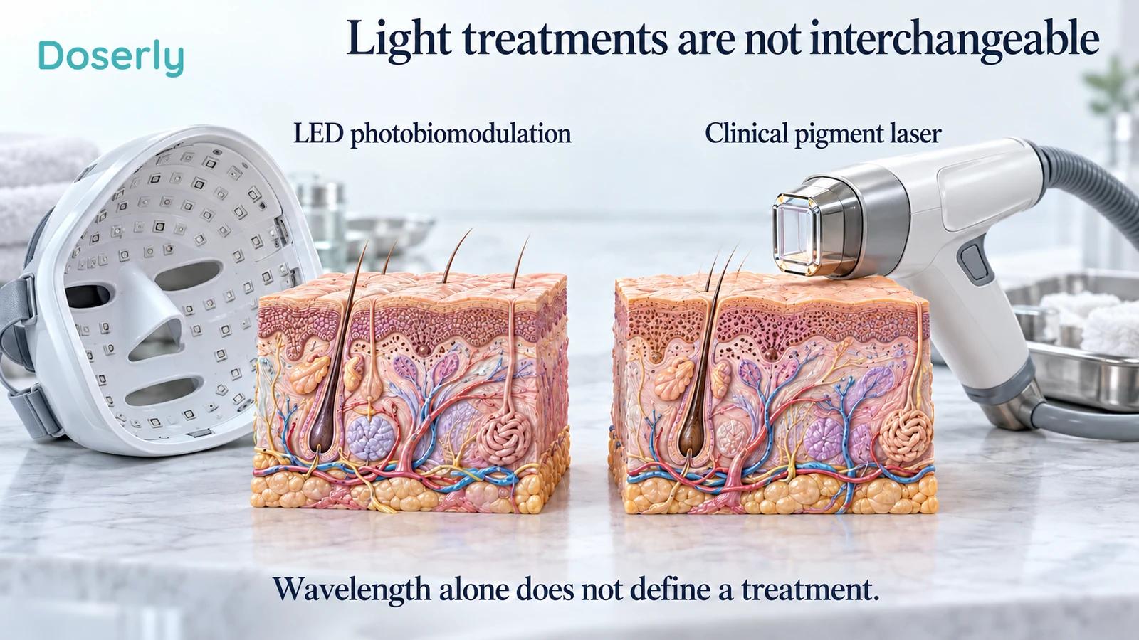
Conceptual equipment comparison illustrates different delivery systems. The skin models do not represent treated patients, and a shared wavelength does not establish equivalent effects.
More aggressive treatment can create more pigment
Trying to remove a stubborn mark can become a cycle of irritation and further discoloration. The possibility is especially relevant when moving from a gentle-sounding device claim to a treatment that produces a stronger tissue response.
In a 2010 randomized split-face study of twenty-nine people, one side received a nonablative 1550-nanometre fractional laser and the other a topical combination. Nine participants developed post-inflammatory hyperpigmentation after two or more laser sessions. Several assessments showed worsening on the laser-treated side, and most participants preferred the topical treatment at six months.
That finding applies to the studied laser protocol, not to all lasers or red LEDs. Its value here is to challenge the assumption that a more intensive light procedure must produce a better pigment result. A treatment intended to improve skin can also provoke a response that complicates the original problem.
This is why warmth, persistent redness or stinging should not be treated as proof that a device is working. If a new routine is making the skin uncomfortable or darker, the useful response is reassessment. Increasing exposure to chase a result can make it harder to identify what caused the change.
A clinician can also distinguish increased pigment from other changes in appearance. Temporary redness, dryness, a new topical product and different lighting can all alter how a patch looks. That distinction helps avoid both overlooking a problem and blaming a device for a change that has another explanation.
Photoprotection remains relevant even when treatment is being studied
The logic of pigment treatment includes limiting the exposures that keep stimulating the condition. A device session does not remove the rest of the day's light exposure or replace a practical protection plan.
A 2025 study in women with skin types III to VI compared SPF50 alone with SPF50 plus an iron oxide-containing foundation over twelve weeks. Both regimens improved several appearance measures. The added formulation produced favorable findings in some melasma outcomes, including colorimetric response and patient-reported quality of life. Most authors were affiliated with the cosmetics manufacturer's research division, and the findings concern those tested formulations.
This does not mean any makeup product provides a known level of visible-light protection. Nor does it mean an SPF number quantifies protection against every wavelength. The study supports considering visible-light protection alongside ultraviolet protection, particularly when pigmentation is the clinical concern.
The AAD's melasma treatment guidance places sun protection and appropriately selected topical treatment within the core plan, with procedures considered for selected patients. Options depend on diagnosis, tolerability and medical circumstances. A new mask should be evaluated in that context, rather than becoming a reason to abandon a working treatment.
A protection plan also needs to be usable. Shade, a hat and a suitable sunscreen formulation serve different practical roles. If a product feels unpleasant or does not blend acceptably, discuss alternatives rather than assuming an impractical routine will become easy through determination alone.
Skin-tone representation is part of the evidence
A trial's inclusion of darker skin is valuable, but it should be described precisely. The studies above range from healthy volunteers with selected phototypes to women from particular clinical populations. They do not provide a complete map of responses across all complexions, pigment disorders, ages or treatment histories.
Fitzpatrick categories describe sun-response patterns; they are not a substitute for a full description of ethnicity or an individual's skin. Two people assigned the same category can have different baseline pigmentation, melasma activity and histories of irritation. A product's statement that it is “for all skin tones” deserves evidence beyond the presence of a few participants in several categories.
It is reasonable to ask how many people similar to you were studied, what adverse changes were actively assessed, and how long participants were followed. If a study reports no pigment complication after a short course, that is a different claim from demonstrating long-term safety during repeated home use.
These questions should improve access to an appropriate treatment, not discourage people with darker skin from seeking care. The useful standard is thoughtful selection with relevant evidence and follow-up, rather than either blanket reassurance or a blanket ban based on complexion.
Track a defined patch, not a vague promise of brightness
Melasma studies use scores such as MASI or modified MASI to summarize aspects of the affected area and darkness. Other tools assess melanin-related color, redness or standardized photographs. They measure related features, but a change in one does not necessarily mean the whole condition has improved to the same degree.
A clinic may use a handheld colorimeter or a calibrated imaging system. At home, the most useful contribution is usually consistency: the same room, angle, camera distance and lighting, without a new filter or coverage product. A photo taken immediately after treatment can be especially difficult to compare if warmth or redness has changed the skin's appearance.
Record what else changed during the same period. A new sunscreen, fewer acne flares, a prescription cream or a change in sun exposure can all affect interpretation. This is not a reason to withhold useful care to create a personal experiment. It is a reason to be honest about what a before-and-after comparison can show.
Agree on what would count as enough improvement to continue. A slightly lower pigment reading, a visibly less prominent patch and greater comfort leaving the house are different outcomes, each worth discussing. The decision should reflect both the skin findings and the burden of the treatment.
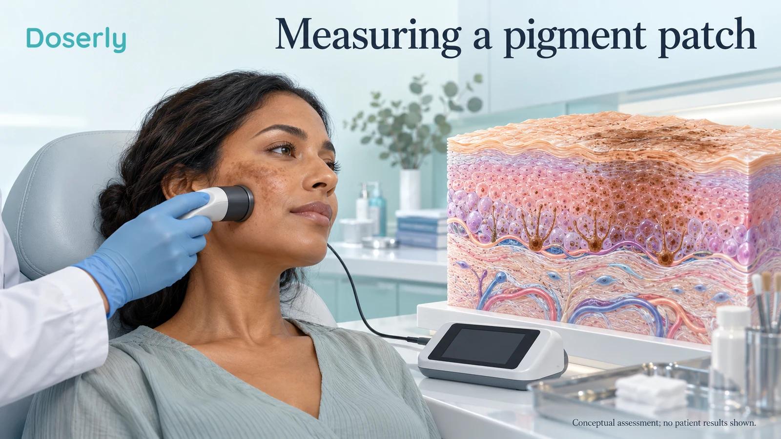
Conceptual pigment assessment illustrates a measurement approach. The person is not a study participant, and no clinical reading or treatment outcome is shown.
Where this leaves a home red-light device
For melasma or persistent dark marks, the evidence supports curiosity about specific light treatments and caution about broad retail promises. The strongest next step is often clarifying the diagnosis and reviewing the existing plan, including ongoing inflammation, light exposure and tolerability.
If a dermatologist considers a device reasonable, ask which study supports that exact approach, whether it tested light alone, and what follow-up would detect benefit or worsening. Keep the distinction between red, amber, near-infrared and clinical laser evidence visible throughout that conversation.
There may eventually be a clearer role for selected photobiomodulation devices in pigment care. Current studies have not settled a universal wavelength, home schedule or maintenance strategy. A useful treatment should improve a problem you have identified without creating a new one, and its claims should be no broader than the people, device and outcomes actually studied.