
Red Light Therapy
October 17, 2026
Red light therapy for dry macular degeneration: what Valeda changes
On this page
For someone living with dry age-related macular degeneration, a treatment intended to improve vision is meaningful news. Much of the conversation around the condition has focused on monitoring, reducing risk, and adapting to changes in sight. Valeda adds a specific light-based treatment to that conversation for a defined group of patients.
It also creates an easy misunderstanding. An authorized ophthalmic device is not evidence that looking into a red light panel helps the eyes. Nor does an improvement on a vision chart mean that age-related macular degeneration has been reversed or that future vision loss has been prevented.
The most useful way to understand Valeda is to keep three questions together: who qualifies under its indication, how much benefit the controlled trials found, and what uncertainty remains about progression and safety. New research published in 2026 makes the evidence more informative, but it does not remove the need for an individual retinal assessment.
Why the diagnosis and stage matter
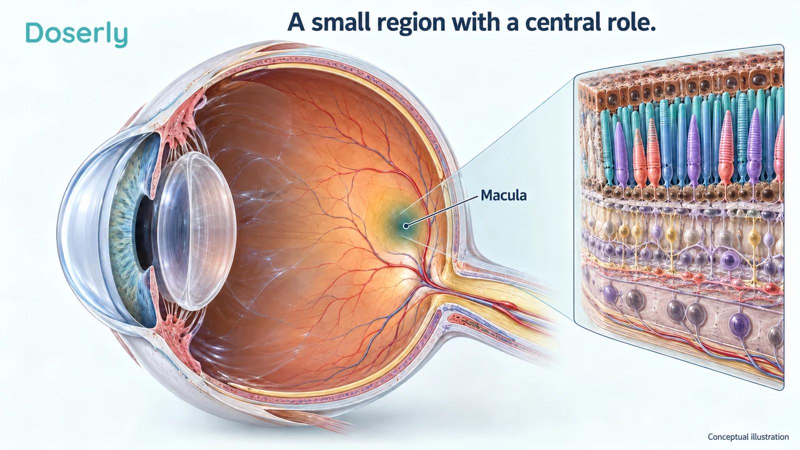
Age-related macular degeneration, usually shortened to AMD, affects the macula, a small region of the retina responsible for detailed central vision. Changes there can interfere with reading, recognizing faces, or seeing details directly ahead. Early disease may cause no noticeable symptoms, while later disease can produce blurred or distorted central vision. The National Eye Institute explains AMD and its symptoms.
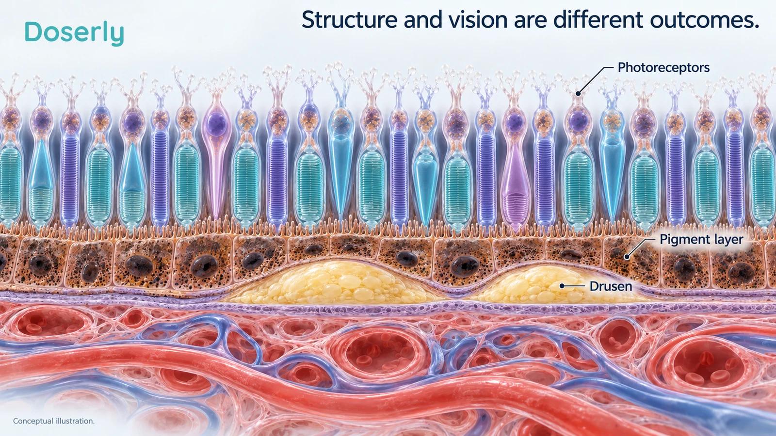
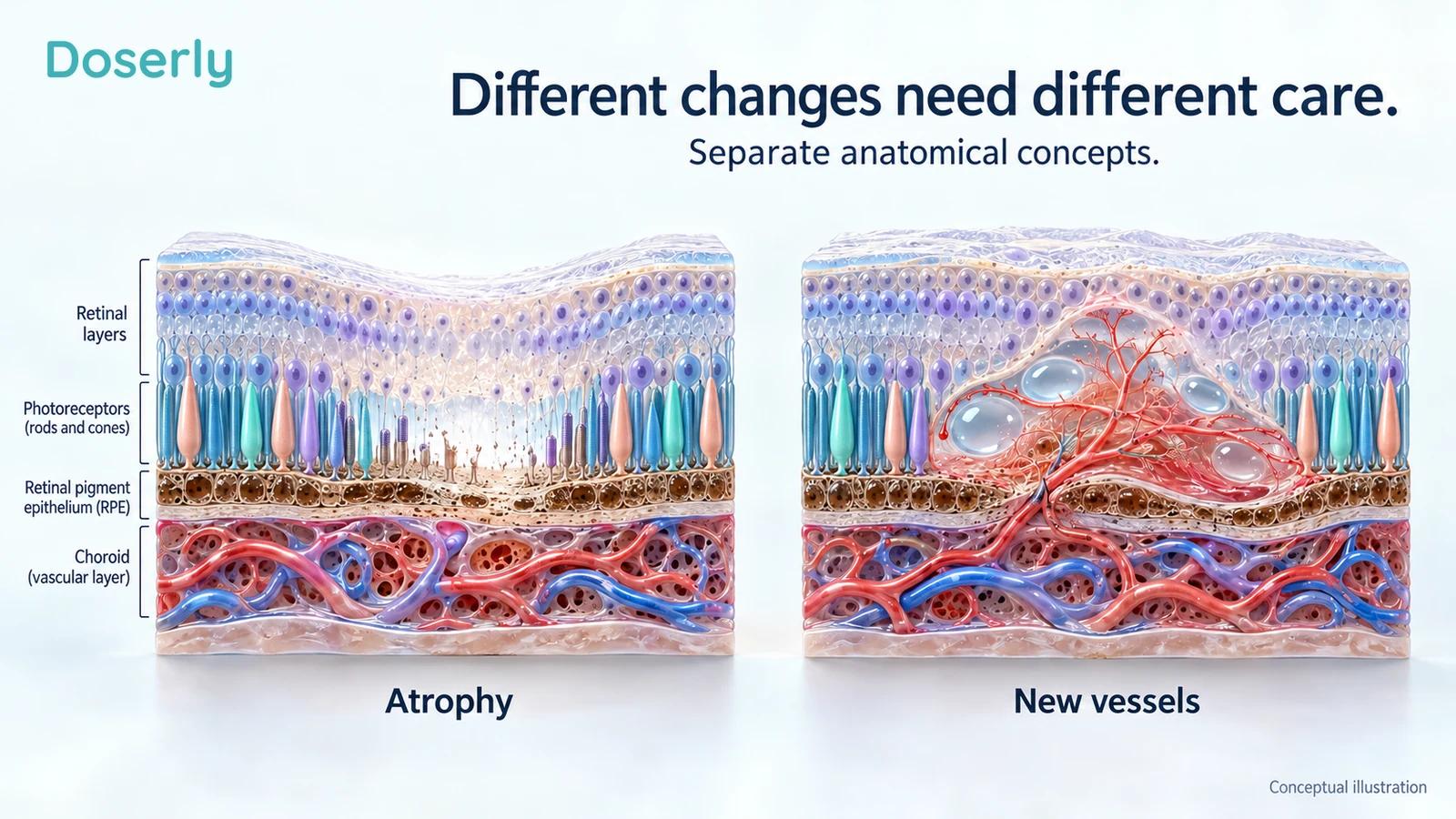
Dry AMD is not a single uniform state. A retina specialist evaluates the type and extent of deposits called drusen, any loss of retinal tissue, and whether abnormal blood vessels are present. Geographic atrophy describes an advanced form of tissue loss. Neovascular, or wet, AMD involves abnormal new vessels and requires its own treatment decisions.
That terminology matters because a study in one stage cannot establish treatment effects in all the others. A person with relatively preserved retinal tissue and modest acuity loss is not in the same situation as someone with extensive central atrophy. Both deserve useful care, but the applicable evidence can differ substantially.
It also matters which eye is being discussed. The two eyes can have different stages, different visual acuity, and different treatment histories. When a clinician describes eligibility, ask whether the explanation applies to the right eye, the left eye, or both. A general diagnosis on a problem list cannot provide that detail.
The FDA indication is narrower than “red light for eyes”
The FDA granted Valeda’s De Novo request on November 4, 2024, establishing it as a Class II prescription device. The FDA database identifies the device and decision. This is a particular authorization with a particular intended use, not an endorsement of every product described as photobiomodulation.
The authorized population has dry AMD and best-corrected acuity between 20/32 and 20/70. The retinal findings must include at least three medium drusen, with diameters greater than 63 and no greater than 125 micrometers; large drusen exceeding 125 micrometers; or geographic atrophy outside the center. Neovascular maculopathy and center-involving geographic atrophy must be absent. The intended benefit is improved visual acuity. These criteria come from the FDA classification order.
Best-corrected acuity means vision assessed with the appropriate optical correction. It is not simply the smallest print someone can read at home without glasses. The retinal criteria also require an examination and imaging interpretation. They cannot be checked by matching symptoms to a website.
This distinction is useful even for someone who already knows they have AMD. “I have dry AMD” begins the conversation; it does not finish the eligibility assessment. Ask the clinician to explain how the measured acuity and retinal findings fit the indication, and what other eye conditions could be contributing to the difficulty seeing.
What the treatment actually is
Valeda is a professional ophthalmic system that delivers selected amber, red, and near-infrared wavelengths: 590, 660, and 850 nanometers. Its optical design, delivery sequence, and clinical controls belong to the tested device. The FDA review describes the system and professional use.
The name photobiomodulation refers to an attempt to influence tissue biology with light. Mitochondrial and cellular stress pathways are proposed explanations, but an attractive mechanism is not the same as a demonstrated clinical benefit. For a person deciding about treatment, the vision and safety results deserve more weight than a diagram of cellular energy.
The evidence also concerns a course of treatment with repeat visits. It should not be imagined as a single session that permanently changes the disease. A clinic can explain the schedule supported by the device labeling, the monitoring involved, and how it would assess whether continuing is worthwhile.
This article does not turn those study schedules into instructions for eye exposure. A household lamp, skincare mask, handheld laser, or general-purpose panel has different intended uses and delivery characteristics. Sharing one wavelength with a studied ophthalmic device does not make its exposure equivalent.
The pivotal trial: a modest average advantage over sham
LIGHTSITE III randomized 100 people, contributing 148 eyes, to Valeda treatment or a sham comparison. The published 13-month analysis reported an average improvement of 5.4 letters in the active group and 3.0 letters in the sham group. The difference attributable to the comparison was therefore about 2.4 letters, with a statistically significant between-group result. The original 13-month report gives both changes.
Keeping both numbers visible is important. Reporting only the active group’s improvement makes the additional treatment effect appear larger than the controlled comparison supports. People in a sham group can improve too, including through measurement variation, repeated testing, and other influences that the comparison helps account for.
This does not make the result meaningless. It makes it specific. The trial detected an average benefit on best-corrected acuity in the studied population. It did not show that every treated eye improved, that normal vision returned, or that all aspects of everyday sight changed to the same degree.
The participants and eyes are also different denominators. A study with 148 eyes is not a study with 148 independent people. When both eyes are included, an analysis needs to account for their relationship. That is one reason careful trial reporting describes the statistical approach rather than presenting the eye count as if it were a larger group of unrelated participants.
What the 2026 two-year publication adds
The 2026 report follows the same LIGHTSITE III trial. Its sham used reduced visible light and omitted near-infrared light, rather than an entirely inactive exposure. At month 21, the estimated acuity changes were 6.2 letters with treatment and 2.4 with sham, an advantage of about 3.8 letters. At month 24, the corresponding changes were 5.6 and 1.3 letters. Treatment had been repeated during the study; these are not two years of benefit after one course. The full two-year report provides the analyses.
It also reported new geographic atrophy in 6.8% of treated eyes versus 24.0% of sham eyes at month 24. This potentially important finding was descriptive and not adjusted for multiple comparisons. It therefore supports further investigation of progression, rather than a settled claim that the device prevents advanced AMD. Twenty participants discontinued over two years. LumiThera and the National Eye Institute supported the study, and several authors were company employees or consultants.
For the person reading about these findings, the central question remains practical: how much would an average difference of several letters matter in the activities that are difficult? A chart score is useful, but it does not directly answer whether someone will read a particular book comfortably or meet a driving requirement. Those outcomes need their own assessment.
Earlier studies explain both the interest and the uncertainty
LIGHTSITE I was a small double-masked, sham-controlled study involving 30 people and 46 eyes. The active group showed an average four-letter improvement immediately after each of two treatment series, with some additional functional and anatomical signals. The researchers emphasized repeat treatment to maintain the observed benefits. The original pilot study describes those findings.
A pilot can establish that an idea deserves a larger test without establishing how reliably it will help in ordinary practice. The responder subgroup in that study should also be interpreted carefully: describing people who improved substantially after treatment is not the same as knowing in advance who will improve.
LIGHTSITE II enrolled 44 people and 53 eyes but was disrupted by the COVID-19 pandemic. Its full report shows a detail that can disappear in brief summaries: at month nine, the main active-versus-sham acuity comparison was not significant. The average changes were approximately 2.3 and 2.0 letters, respectively. An analysis restricted to participants completing the entire treatment schedule also did not establish a significant between-group advantage. The full LIGHTSITE II results make these comparisons explicit.
Interrupted treatment, missed visits, and the small sample complicate interpretation. They do not justify silently turning a within-group improvement into a proven advantage over sham. Taken together, the LIGHTSITE studies show a developing evidence base with a positive pivotal result, rather than three identical demonstrations of a large effect.
Other devices and research groups broaden the picture
A 2024 randomized study of the EYE-LIGHT system included 76 patients and 152 eyes in its preliminary analysis. This device uses a different wavelength combination from Valeda. At four months, 20.3% of treated eyes versus 8.9% of sham eyes gained at least five letters. Central retinal thickness did not differ significantly between groups, while a drusen-volume measure favored treatment. The original report describes the device-specific results.
The finding is relevant to the wider field, but it does not make the devices interchangeable. Safety, tolerability, and compliance were the primary focus of that preliminary report; the functional and anatomical findings were additional outcomes. Four months also cannot establish durable protection against progression.
A 2026 multicenter report from an overlapping investigator group studied 138 eyes from 78 patients over 12 months. Its primary outcome was drusen volume, which favored PBM. Visual acuity was secondary: the reported adjusted between-group difference was 3.75 letters, with a 95% confidence interval from 1.16 to 6.34. The original abstract and publisher disclosure describe the findings.
The full methods were not accessible for this review, so the exact cohort relationship to the earlier report could not be independently established; they should not be counted as clearly independent replications. Some authors disclosed speaking fees from a device company. The result adds useful evidence, while leaving longer-term progression and device-specific applicability open.
An independent prospective PBM4AMD study followed 38 people who completed treatment. Several visual measures improved initially but weakened by six months, and two participants developed macular neovascularization. There was no randomized sham comparison to establish causation or treatment-attributable risk. The original study reports the changing pattern over time.
A separate 2026 retrospective series of 30 patients and 47 eyes found a roughly three-letter average acuity improvement, with limited short-term structural change. Without a concurrent control group, it cannot separate treatment effects from other explanations. That clinical series is useful observational context. It does not have the evidentiary role of another randomized trial.
Retinal structure and visual function are different outcomes
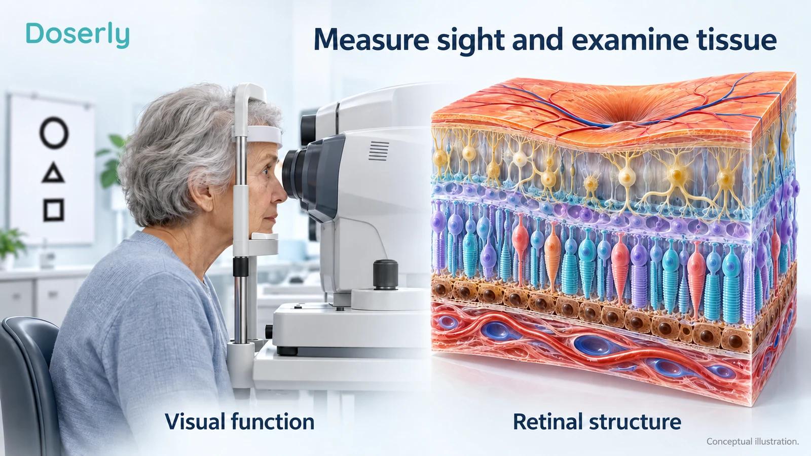
Drusen volume is an anatomical measurement. Visual acuity measures recognition of detail under standardized conditions. Contrast sensitivity, reading performance, and low-light vision describe other aspects of function. A treatment can affect these measures differently, which is why a paper should not compress them all into the phrase better eye health.
The newer multicenter trial illustrates this distinction particularly well: its main question concerned drusen, while acuity was a secondary question. The retrospective series provides another example, with a functional change despite limited structural change. Neither pattern should be rewritten as evidence that retinal cells were regenerated.
For a reader, a helpful habit is to translate every claim into its actual measurement. “Less drusen” means an imaging measurement changed. “More letters” means a standardized acuity score changed. “Less progression” requires disease events assessed over a suitable period. These are related clinical concerns, but one cannot substitute for another simply because all involve the retina.
The same care applies to illustrations. A healthy-looking retinal model can explain anatomy; it cannot show what happened to a participant unless it is an actual appropriately presented clinical image. The figures here are teaching models, not before-and-after scans or representations of measured treatment effects.
Safety includes watching for wet AMD
The FDA review notes that progression to neovascular AMD occurred more often in the active arm of the pivotal study. It could not determine whether this reflected differing baseline risks, chance, a treatment effect, or a combination. It specifically calls for close monitoring when the fellow eye already has neovascular AMD. The same review highlights limited evidence outside the studied population, including no treated non-White participants, and precautions concerning relevant photosensitivity and light-triggered neurological disorders. The FDA’s benefit-risk assessment explains these uncertainties.
This is more informative than either claiming the treatment causes wet AMD or saying that a favorable overall safety description means there is nothing to monitor. A small study can identify a concern without determining its cause. Professional follow-up is part of interpreting that concern, not an optional addition to an otherwise complete treatment.
New distortion, especially straight lines appearing wavy, warrants prompt contact with an eye-care professional rather than waiting for the next light session. The National Eye Institute identifies this as a warning sign. A change in symptoms should lead to assessment, not an assumption that the treatment needs more time.
Bring a complete medication and supplement list to the appointment. The clinician can evaluate the relevant device precautions in the context of the actual products, conditions, and eye findings. Broad online lists of supposedly safe or unsafe combinations cannot replace that assessment.
Valeda does not replace the rest of AMD care
AREDS2 supplements have a separate evidence base and a stage-specific role. The National Eye Institute describes their use for intermediate AMD and selected people with late AMD in one eye; they are not a general way to prevent AMD from starting. The formulation also matters, particularly the avoidance of beta-carotene in people who smoke or previously smoked. NEI’s AREDS2 guidance explains the intended population and cautions.
The original AREDS2 trial enrolled 4,203 people at risk of advanced AMD. Its primary analyses did not show an additional significant benefit from adding lutein and zeaxanthin, omega-3 fatty acids, or both to the background AREDS formulation. The beta-carotene safety findings helped support replacing that ingredient with lutein and zeaxanthin. The original randomized trial describes this more nuanced result.
Longer-term follow-up of the same trial population subsequently supported lutein and zeaxanthin as an appropriate replacement, while retaining the concern about beta-carotene and lung cancer. The follow-up was observational after the randomized treatment period, with AREDS2 supplements offered to all participants. The ten-year follow-up report explains that design.
For background on one of these ingredients, Doserly’s lutein guide discusses its own evidence. A single ingredient is not automatically equivalent to the studied combination, and neither supplements nor light treatment should be treated as interchangeable solutions. The useful question is how each fits the diagnosed stage and the rest of the care plan.
The same principle applies if the disease changes. Care appropriate for one stage may need to change with new findings. An earlier decision to try Valeda is not a commitment to keep the same plan regardless of what subsequent examinations show.
Support for daily vision remains worthwhile
A treatment decision should leave room for help with the activities that are difficult now. Low-vision services address how a person uses remaining vision, rather than depending on an experimental treatment to change the underlying disease. This is a different clinical goal from gaining letters on an acuity chart.
The LOVIT II randomized trial enrolled 323 veterans with macular diseases and compared low-vision devices alone with devices plus therapist-led rehabilitation. The added rehabilitation improved several functional measures, including reading, with the clearest added benefit among participants with poorer starting acuity. There was no significant added benefit in the milder acuity subgroup. The sample was 97% male, which limits its representation of the wider AMD population. The original rehabilitation trial describes these results.
This was not a comparison with Valeda, and its participants were not selected according to Valeda’s indication. It nevertheless shows why daily function deserves a place in the conversation. An intervention can help a person perform tasks without demonstrating retinal regeneration or slowing the disease.
Bring concrete examples to an appointment: difficulty reading medication labels, recognizing a familiar face across a room, or following a recipe. These examples help the clinician understand what a chart score means in your life. They also provide a basis for discussing appropriate support while the medical treatment plan is being evaluated.
If you do not meet the device indication, that does not mean there is nothing to discuss. Ask what care is relevant to your actual findings and which services could make the most troublesome tasks easier. If you do qualify, those same questions remain worthwhile. Device eligibility is one part of care, not a complete description of your needs.
Questions that make a treatment discussion more useful
Start with eligibility: which eye fits the authorized criteria, and what finding establishes that? Ask for the measured acuity and a plain-language explanation of the retinal images. This helps distinguish a treatment intended for the condition you have from a general offer based on the word AMD.
Then discuss the expected outcome. Is the goal a modest acuity improvement, a change in a particular daily task, or something else? Ask how progress will be measured and how uncertainty about future progression will be explained. A realistic conversation can acknowledge a possible benefit without promising restored sight.
The practical commitment deserves equal attention. Ask about the full course of visits, planned reassessment, transportation, and the total expected financial burden. Coverage and local availability can change, so obtain those details directly from the clinic and insurer. A treatment that is manageable in principle may be difficult to sustain if every visit requires substantial travel or help from family.
Finally, agree on what would prompt a change of plan. That might include new symptoms, an examination finding, difficulty attending, or an absence of worthwhile benefit. Defining the review process in advance makes it easier to make a considered decision later, rather than feeling obliged to continue because a course has already begun.
Valeda changes the dry-AMD conversation by offering a tested, authorized option for selected eyes. The human evidence supports a modest average visual-acuity benefit with a specific clinical device, while leaving important questions about durability, progression, and broader applicability. Its value is best assessed through the details of the individual eye and the controlled evidence, with ongoing retinal care at the center of the decision.