
Peptides
October 16, 2026
KPV and alpha-MSH: related does not mean interchangeable
On this page
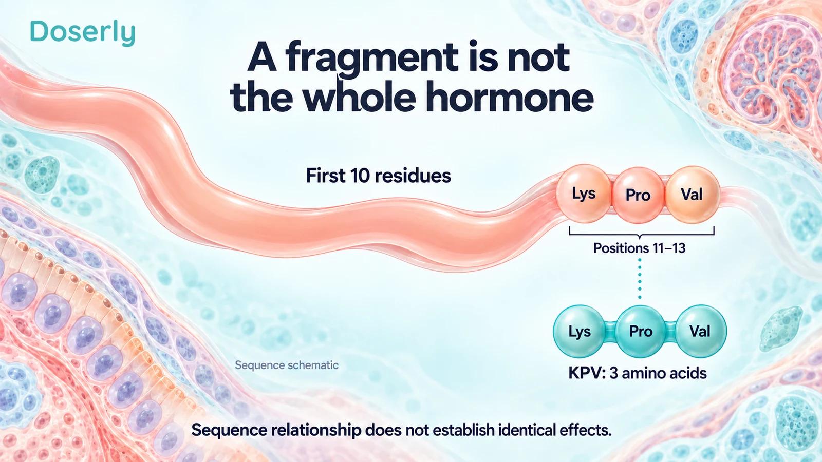
KPV is often introduced as a fragment of alpha-melanocyte-stimulating hormone, usually written alpha-MSH or α-MSH. That relationship is real and useful. KPV corresponds to the lysine–proline–valine sequence at positions 11–13, the end of the longer peptide. It helps explain how KPV entered the research literature.
It does not make KPV a miniature replacement for every action of alpha-MSH. Shared amino acids do not establish identical receptor interactions, delivery, dose, safety or clinical benefit. In fact, some of the experiments that make KPV interesting specifically show differences between the fragment and other melanocortin peptides.
The most accurate account is therefore more nuanced than either “the same hormone, but smaller” or “completely unrelated.” There is a sequence relationship and there are experimentally observed functional distinctions. The KPV guide gives a broader introduction; this article explains why the distinction matters when reading claims about inflammation, pigmentation and peptide products.
What the fragment relationship means
Alpha-MSH is commonly described as a 13-amino-acid peptide. KPV names three amino acids in order: K for lysine, P for proline and V for valine. Research papers may call this sequence MSH(11–13), referring to its position within the parent peptide. Early experimental work explicitly investigated the activity of this terminal fragment. Hiltz and Lipton’s original study.
A sequence relationship is a statement about composition. It does not tell us whether the isolated fragment behaves like the intact parent in every setting. Interactions with biological targets depend on the molecule that is actually present, not just whether a familiar sequence occurs somewhere within it.
Even the notation can hide important differences. A paper may study a terminally modified peptide, a particular salt, an amino-acid substitution or a linked form containing more than one peptide unit. “KPV-related” can be a useful broad category for discussing research, but it is too broad to establish that two preparations are interchangeable.
Why researchers studied the short sequence
The early terminal-fragment experiments asked whether a portion of alpha-MSH retained measurable biological activity. In a 1989 mouse study, researchers compared effects of the terminal peptide on chemically induced ear swelling with comparison treatments and reported reduced swelling. The experiment helped support further investigation of short related peptides. Original mouse study.
The setting matters. Ear swelling in a mouse model is a defined experimental outcome; it is not a demonstration of treatment for human eczema, inflammatory bowel disease or a general state called “systemic inflammation.” Nor does a comparison within one experiment establish equivalence between an isolated fragment and the parent hormone across other tissues.
This is the productive reason to study fragments: they can help identify which structural features contribute to particular effects. The result is a more detailed hypothesis, not permission to carry all of the parent molecule’s properties into a product description for the fragment.
Similar inflammatory findings can involve different signaling
In a 2001 study, investigators compared alpha-MSH, its first ten residues and its terminal three residues in macrophage-like cells. The peptides affected inflammatory measurements, but their receptor-binding and cyclic-AMP responses differed. The terminal fragment did not compete in the reported MC1 receptor-binding assay or stimulate cyclic AMP as the longer peptides did. The authors proposed more than one mechanism. Mandrika and colleagues.
Cyclic AMP is an intracellular signaling molecule. A change in it can help researchers investigate how a response occurs. Its absence in a particular assay does not mean that nothing happened in the cell; its presence does not by itself prove a useful treatment effect.
That is why “both reduced an inflammatory measurement” is not the same as “both work identically.” A common endpoint can be reached through different biological events. The study’s comparisons are valuable precisely because they separate a shared observation from an assumed shared mechanism.
For a consumer claim, the boundary is important. One cannot infer every receptor-mediated effect of alpha-MSH from an inflammatory response to KPV in a cell system. The experiment needs to establish the specific interaction and response being discussed.
A mouse comparison also found functional differences
Getting and colleagues compared KPV with alpha-MSH and other melanocortin-related peptides in crystal-induced peritonitis in mice. KPV reduced accumulation of inflammatory cells, and its effect persisted under tests that challenged conventional melanocortin-receptor explanations. In parallel cell work, however, KPV did not reproduce all the responses produced by the other peptides. Original 2003 study.
The authors interpreted the results as evidence that the terminal fragment’s anti-inflammatory effect differed from the core melanocortin peptides. This is stronger support for distinguishing the compounds than a generic statement that they belong to the same family.
It is still a model-specific inference. The experiment does not settle every potential target of KPV in every tissue, and it does not establish human efficacy. A careful explanation preserves both points: the findings challenge a simple same-mechanism story, while leaving further questions open.
Why “KPV never involves melanocortin receptors” is too absolute
A 2004 study examined signaling in human keratinocyte systems and engineered cells expressing MC1 receptor. The investigators did not detect elevated cyclic AMP in the keratinocytes under the tested conditions, but they observed calcium responses with important experimental qualifications. In the receptor-expressing engineered cells, alpha-MSH and KPV peptides increased intracellular calcium. Elliott and colleagues.
That finding complicates a blanket claim that all KPV effects are entirely independent of melanocortin receptors. It does not prove that every proposed KPV effect uses MC1 receptor either. Different cell systems, conditions and signaling readouts need to remain visible rather than being reduced to a universal yes-or-no slogan.
The distinction between binding, signaling and clinical outcome is especially useful here. An assay may examine competition with a labeled ligand, a change in cyclic AMP or a change in intracellular calcium. These are related investigative tools, not identical measurements. A negative result in one does not automatically settle all the others.
This nuance is relevant to pigmentation claims. The studies described here do not establish that a commercial KPV product has a particular effect on human skin color, or that it shares the full parent hormone’s effects. They also do not establish a comprehensive absence of such effects in people. Human product-specific evidence would be needed for those claims.
PepT1 adds a different kind of mechanism
The KPV gut literature introduces PepT1, a transporter for small peptides. In intestinal epithelial and immune-cell experiments, researchers connected PepT1-mediated uptake with KPV’s effects on inflammatory signaling. They also studied oral delivery in mouse colitis models. Dalmasso and colleagues.
Transport and receptor signaling are different questions. A transporter can move a substance across a membrane. A receptor can initiate a signal following an interaction. Identifying a route into a cell does not necessarily identify the final intracellular target, and neither finding alone proves treatment benefit.
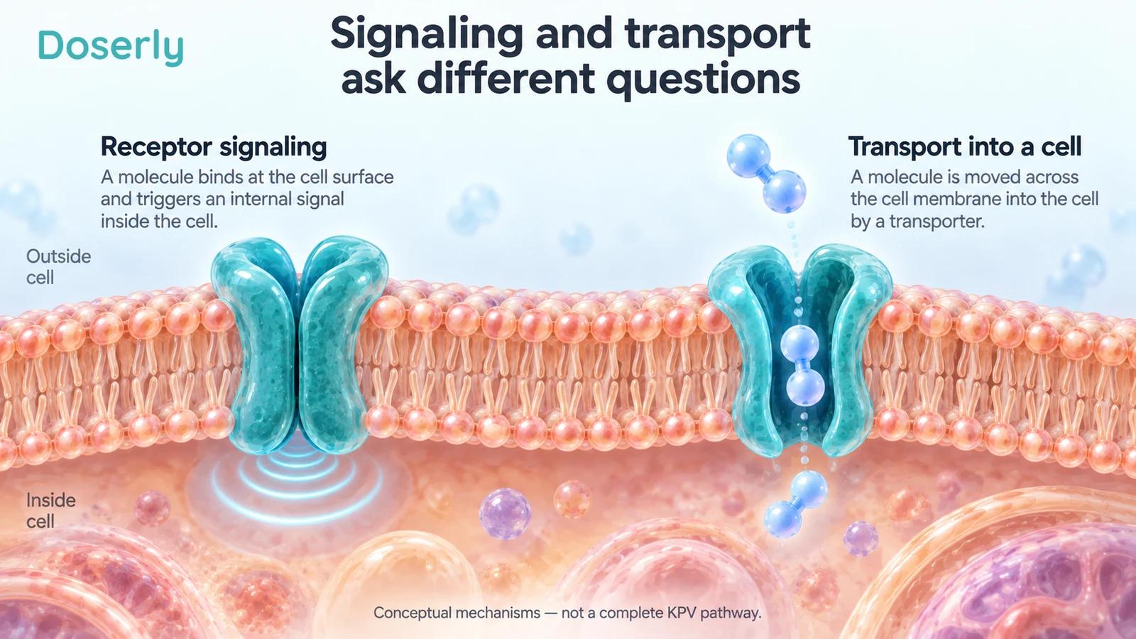
This helps explain why a short peptide’s research story can differ from that of its parent. Removing most of the sequence may change the relevant questions about binding and transport. Researchers have to investigate the resulting molecule, rather than assuming that the parent’s pharmacology simply survives in a smaller package.
Related clinical trials cannot be assigned to KPV
The naming issue becomes particularly consequential when human trials are cited. A randomized ulcerative-colitis study evaluated K(D)PT as an add-on to baseline treatment. Its abstract reports preliminary results with a high placebo response and additional analyses. It was not a trial of KPV. Original K(D)PT trial.
The original registry identifies the ingredient as L-lysyl-D-prolyl-L-threonine acetate. That means both the specified proline stereochemistry and the final amino acid differ from the simple KPV sequence. Calling it merely another spelling of KPV would lose information essential to interpreting the trial. Trial ingredient record.
The same principle applies to studies of full-length alpha-MSH or another analog: they can inform a research rationale without establishing a clinical result for KPV. A reader should be able to follow the name in a product claim through to the substance actually administered in the cited study.
“Related to a natural hormone” does not establish product safety
The FDA’s July 2026 KPV briefing reported no identified human clinical studies or exposure data for KPV free base or acetate by any route, and no human pharmacokinetic or pharmacodynamic studies. The document also discussed uncertainties in chemical identity and product characterization. It was an advisory-committee briefing, not a declaration that KPV had completed drug approval. FDA briefing.
The sequence’s relationship to an endogenous peptide does not fill those gaps. A manufactured preparation introduces questions about identity, formulation, impurities, route and exposure. Those questions remain even when the amino acids themselves are familiar.
Likewise, “non-hormonal” is not a complete safety argument. The meaningful question is what the administered material does under the proposed conditions of use. A label cannot substitute for studies of the actual preparation, dose range, population and duration.
A practical way to check a peptide-family claim
First, identify the exact molecule. Is the paper about full alpha-MSH, KPV, a modified terminal fragment, K(D)PT or a dimer? If the name changes between the claim and the citation, the explanation should say why that other evidence is relevant and what it cannot establish.
Second, identify the model. A human cell line, mouse inflammatory model and clinical trial in patients answer different questions. Human-derived keratinocytes, as used in the 2004 signaling study, do not turn a signaling experiment into a clinical skin-treatment study.
Third, identify the measurement. Receptor binding, calcium signaling, cytokine release, tissue inflammation and symptom relief should not be treated as synonyms. A scientifically useful summary names the observed response rather than replacing it with an all-purpose phrase such as “balances immunity.”
Finally, check the scope of the conclusion. Evidence that two peptides differ in one signaling assay is not evidence that one is globally safer or better. Evidence that a fragment has an effect in an animal model is not evidence that it reproduces all the parent hormone’s actions in humans.
KPV’s relationship to alpha-MSH is a reason to ask sharper questions about structure and function. The original studies make that relationship more interesting, not less: they show why a fragment can deserve investigation in its own right. Keeping its identity separate from the parent hormone is essential to understanding what has actually been learned.
Sources
- Hiltz and Lipton, terminal-fragment mouse experiment.
- Mandrika et al., differing signaling mechanisms.
- Getting et al., core and terminal melanocortin peptides.
- Elliott et al., human keratinocyte signaling.
- Dalmasso et al., PepT1-mediated KPV uptake.
- Kucharzik et al., K(D)PT clinical trial.
- K(D)PT registry ingredient identity.
- FDA, July 2026 KPV briefing.