
Peptides
October 16, 2026
DSIP and deep sleep: what an EEG can actually establish
On this page
Deep sleep is a real physiological state, not simply a description of an especially satisfying night. Researchers identify it using patterns of brain activity recorded during sleep. That makes it measurable—but it doesn't mean every change in a brain-wave recording represents better sleep or better health.
This distinction matters for delta sleep-inducing peptide, or DSIP. The name suggests a connection with the slow electrical rhythms associated with deep sleep. Human studies, however, have not established that DSIP reliably increases restorative sleep in people with insomnia. Some findings involve lighter sleep stages, and one frequently relevant EEG experiment was conducted during surgical anaesthesia rather than ordinary sleep.
We'll look at what the recordings mean, what the DSIP experiments actually measured and why a useful treatment needs to do more than change an electrical signal. For the compound's broader background, see our DSIP guide.
What an EEG records
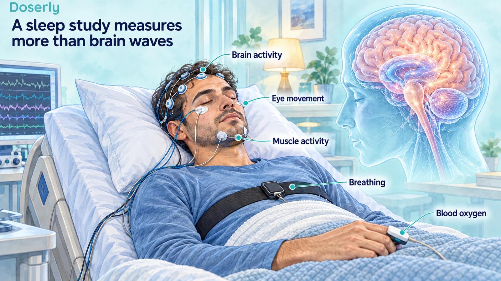
An electroencephalogram, or EEG, uses scalp electrodes to record electrical activity arising from the brain. It gives researchers a changing signal over time, which can be examined for patterns associated with wakefulness and sleep. It does not directly read thoughts, count repaired brain cells or measure how refreshed someone will feel the next morning.
In a full sleep study, called polysomnography, EEG is combined with other measurements. These can include eye movements, muscle activity, breathing, heart rate and blood oxygen. The additional signals help distinguish sleep states and identify disturbances that a brain-wave trace alone would miss. NHLBI explanation of sleep studies
For example, a clinician assessing fragmented sleep may need information about breathing interruptions as well as sleep stages. Recording more slow activity would not by itself establish that those interruptions had resolved. A measurement becomes clinically useful when it answers the problem being investigated.
Deep sleep is one stage in a changing night
Non-REM sleep includes stages N1, N2 and N3. N3 is often called deep sleep or slow-wave sleep. People generally spend more time in it earlier in the night, while REM sleep becomes more prominent later. Sleep architecture is the pattern and timing of these stages across the night. NHLBI sleep-stage overview
The word “deep” can make the other stages sound inferior. They are not failed attempts at deep sleep. They are components of the normal sleep cycle, with different physiological characteristics. A treatment should not be judged on an assumption that the best possible night contains the largest possible proportion of one stage.
Age also affects sleep architecture. The NIH overview describes a decline in slow-wave sleep across adulthood. That does not mean an older adult should diagnose a peptide deficiency from a low deep-sleep estimate. It means that a measurement needs an appropriate clinical and age-related context.
Delta activity and minutes of deep sleep are different quantities
A researcher can analyze the amount of electrical activity in a frequency range, or classify intervals into sleep stages and count their duration. Those approaches are related but answer different questions. An increase in a signal characteristic does not automatically tell us how many additional minutes someone spent in a particular stage.
The denominator matters too. Consider a hypothetical night with 60 minutes of deep sleep out of six total hours: deep sleep occupies about 17% of sleep time. If total sleep falls to five hours while deep sleep stays at 60 minutes, the percentage rises to 20%. The percentage improved numerically, but no extra deep-sleep minutes occurred and total sleep decreased. These are arithmetic examples, not DSIP study results.
This is why a claim such as “increased deep sleep” needs a definition. Was the measured change in minutes, percentage, electrical power or an algorithm's estimated stage? Was it compared with placebo, an earlier night or a reference range? Without those details, the headline is incomplete even if the underlying measurement is technically accurate.
What the controlled insomnia study found
Monti and colleagues' 1987 study used polysomnography to assess intravenous DSIP in chronic insomnia under blinded crossover conditions. Some measures moved in a favorable direction, but reductions in awakenings, sleep latency and wake time did not reach significant differences from baseline or placebo. Monti trial
The distinction between stages was especially important. Slow-wave sleep and REM sleep did not change. Increased non-REM sleep was related to stage 2, and differences between DSIP and placebo in stage 2 and total non-REM sleep were already present at baseline. The authors regarded the overall improvement as having little clinical significance.
That finding does not prove that DSIP can never affect a brain signal. It directly limits a more specific claim: this controlled insomnia experiment did not demonstrate an increase in deep sleep. Describing its stage 2 result as “more restorative delta sleep” would substitute a different outcome for the one measured.
It also shows why it helps to read the sleep-stage breakdown rather than only the total. An intervention might alter time spent in one stage while leaving another unchanged. If the claim concerns N3, a result involving N2 cannot fill the gap.
What about improved sleep efficiency?
A separate 1992 double-blind study enrolled 16 people with chronic insomnia. Participants spent five nights in the laboratory and received DSIP or placebo before the final three. Some objective results favored DSIP, including sleep efficiency and time to fall asleep. Yet the authors described the effects as weak, partly related to incidental change in the placebo group, and unlikely to provide a major therapeutic benefit. Subjective sleep quality did not improve. Bes and colleagues
Sleep efficiency is the fraction of time in bed spent asleep. It can be useful, but it does not identify which stage accounts for the sleep or whether the person feels better. A higher efficiency value is therefore neither a direct deep-sleep measure nor a complete assessment of treatment success.
The difference between objective and subjective findings is informative. Laboratory recordings and patient experience can diverge without either being meaningless. They measure different aspects of a night. A strong insomnia trial should plan how to interpret both, rather than treating whichever looks favorable as the only valid outcome.
The anaesthesia study asks another question
In a 2009 experiment, 24 women undergoing surgery were randomized to saline or DSIP. Twelve received saline; the other 12 were allocated among three intravenous DSIP doses. Researchers measured EEG, heart-rate variability and the bispectral index, or BIS, a processed EEG measure used to help assess anaesthetic depth. Original anaesthesia report
The participants received propofol for induction and isoflurane to maintain anaesthesia. Under those conditions, the lowest DSIP dose was associated with reduced delta activity and increased BIS, together with other changes. The authors interpreted the pattern as lighter anaesthesia, the opposite direction from their hypothesis that DSIP would deepen it.
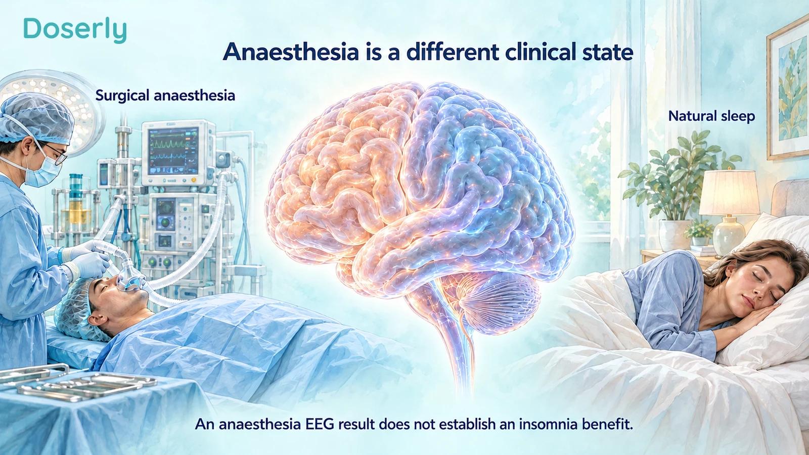
This is a small surgical study, not a test of insomnia treatment. It cannot tell us whether someone would obtain more deep sleep at home, feel more rested or function better the next day. The result is useful precisely because it demonstrates that DSIP's name does not predict the direction of every EEG response in every setting.
Why anaesthetic depth is not sleep quality
During the surgical experiment, EEG activity was measured in the presence of other potent medicines and the circumstances of anaesthesia. During an insomnia experiment, the question concerns a person's ability to initiate and maintain natural sleep. Even if some electrical features overlap, the treatment goals and interpretation differ.
A BIS change is not a scale of how restorative a night has been. Nor can an altered anaesthesia measurement be relabeled as evidence of overnight recovery. The study's endpoint remains anaesthetic depth, and its small dose groups further limit what can be inferred about different doses or settings.
The same principle applies to other physiological measurements in the report. Heart-rate variability describes variation in the intervals between heartbeats. A change can help characterize a physiological response, but does not on its own establish better sleep, lower anxiety or improved health. Those outcomes need to be studied directly.
What would make an EEG finding convincing?
A useful DSIP sleep study would specify the electrical or staging outcome before analyzing the recordings. It would explain how recordings were scored, whether scorers were unaware of treatment assignment and whether the measurements were comparable across nights. These are proposed requirements for interpreting a future study, not claims that a completed DSIP trial met them.
It would also include an appropriate control group. Sleep varies across nights, and becoming accustomed to a laboratory can change the experience. A baseline night provides context but does not necessarily predict what would have happened on every later night without treatment.
Finally, the study would connect the physiological finding to the clinical question. If the proposed benefit is better daytime function, researchers should measure daytime function. If it is fewer prolonged awakenings, those awakenings should be counted. An EEG change could help explain a demonstrated benefit; it should not be used to invent one that was never observed.
What should a person do with a deep-sleep number?
Start by identifying its source. A laboratory EEG-based sleep study, a clinical activity monitor and a consumer wearable may all present sleep information, but they do not collect the same signals. A number labeled “deep sleep” should not be assumed to be a direct EEG measurement unless the device and method actually support that description.
Then put the number alongside symptoms. Persistent difficulty sleeping, substantial daytime sleepiness, witnessed breathing pauses or other concerns deserve a clinical discussion based on the whole pattern. NIH describes sleep testing as a tool for diagnosing several sleep disorders, with the choice of testing determined by the clinician. NHLBI sleep studies
For chronic insomnia, established care includes multicomponent cognitive behavioral therapy for insomnia, which AASM strongly recommends. Its purpose is to treat the disorder and its impact, rather than maximize a single sleep-stage score. AASM guideline
The practical message from DSIP research is specific: brain activity can be measured, but a changed recording is not automatically better sleep. The controlled insomnia findings did not establish a robust deep-sleep benefit, and the anaesthesia experiment belongs to a different clinical question. A worthwhile treatment should help the person, with measurements that show what improved and at what cost.
Sources
- NHLBI, sleep phases and stages.
- NHLBI, sleep studies.
- Monti and colleagues, DSIP and polysomnographic sleep outcomes, 1987.
- Bes and colleagues, objective and subjective insomnia outcomes, 1992.
- DSIP, EEG and bispectral index during isoflurane anaesthesia, 2009.
- AASM, behavioral and psychological insomnia treatment guideline, 2021.